Review
. 2011:80:973-1000.
doi: 10.1146/annurev-biochem-061609-165311.

STIM proteins and the endoplasmic reticulum-plasma membrane junctions

Affiliations
Review

STIM proteins and the endoplasmic reticulum-plasma membrane junctions

Silvia Carrasco et al. Annu Rev Biochem. 2011.

Abstract

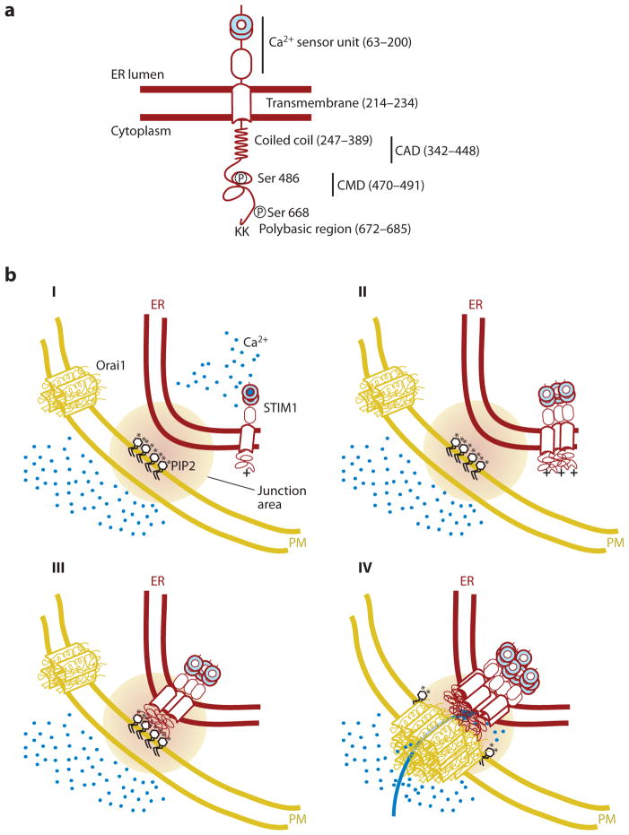
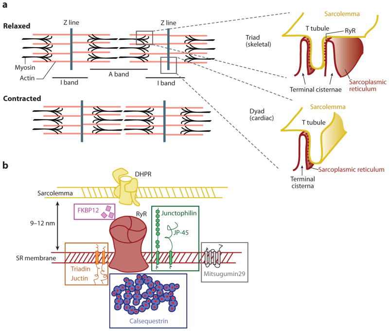
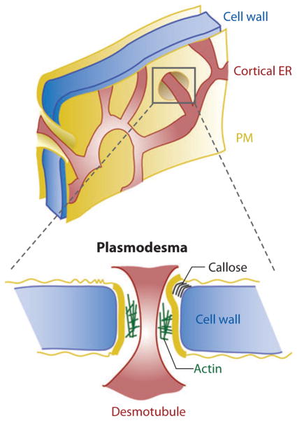
Eukaryotic organelles can interact with each other through stable junctions where the two membranes are kept in close apposition. The junction that connects the endoplasmic reticulum to the plasma membrane (ER-PM junction) is unique in providing a direct communication link between the ER and the PM. In a recently discovered signaling process, STIM (stromal-interacting molecule) proteins sense a drop in ER Ca(2+) levels and directly activate Orai PM Ca(2+) channels across the junction space. In an inverse process, a voltage-gated PM Ca(2+) channel can directly open ER ryanodine-receptor Ca(2+) channels in striated-muscle cells. Although ER-PM junctions were first described 50 years ago, their broad importance in Ca(2+) signaling, as well as in the regulation of cholesterol and phosphatidylinositol lipid transfer, has only recently been realized. Here, we discuss research from different fields to provide a broad perspective on the structures and unique roles of ER-PM junctions in controlling signaling and metabolic processes.


Figures

Figure 1
Figure 1
Schematic representation of the STIM1 translocation process to endoplasmic reticulum (ER)–plasma membrane (PM) junctions following a decrease in ER Ca2+ levels. (a) Main regulatory domains of STIM1. Numbers in parenthesis depict domain-boundary residues in human STIM1. (b) Induced coupling of STIM1 and Orai1 at the ER-PM junction: (I) At high basal Ca2+ (blue circles) in the ER, STIM1 is present as a monomer or dimer relatively uniformly distributed in the ER. Orai1, likely a tetramer, is uniformly present in the PM and is inactive when not bound to STIM1. (II and III) When Ca2+ is depleted in the ER, Ca2+ dissociates from the STIM1 EF-SAM domain, changes its conformation, thereby inducing protein oligomerization, generating a bigger cluster of positively charged amino acids (plus sign), which is now sufficient to interact with PIP2 (black; phosphate group represented as an asterisk) and other negatively charged lipids in the PM. (IV) STIM1 localization to the ER-PM junction, together with an oligomerization-mediated conformational change of STIM1, triggers binding and activation of Orai1, further triggering Ca2+ entry across the PM. CAD, CRAC activation domain; CMD, CRAC modulatory domain.
Figure 2
Figure 2
Schematics of the endoplasmic reticulum (ER)-plasma membrane (PM) junction in striated muscle. (a) The sarcomer, the contraction unit of the striated muscle, as observed by electron microscopy. Actin is shown in pink, and myosin in black (with barbed ends). The inset depicts triad and dyad muscle ER-PM junctions in their common localization along the sarcomer. At these junctions, the terminal cisternae of the sarcoplasmic reticulum (the muscle ER) and the T tubule of the sarcolemma (the muscle PM) are closely linked. The small dots with more intense color in the terminal cisternae represent the ryanodine receptor (RyR), clearly observable as small protrusions by electron microscopy (“feet structures”). (b) List of selected proteins localized to triad- or dyad-type ER-PM junctions. DHPR, dihydropyridine receptor; SR, sarcoplasmic reticulum.
Figure 3
Figure 3
Schematics of endoplasmic reticulum (ER) distribution in yeast. (a) Yeast ER can be separated into sheet-like perinuclear ER and cortical ER structures, connected by ER tubules. The cortical ER is close to the plasma membrane (PM) and forms numerous ER-PM junctions, shown as more intense colored patches. (b) Model of the difference between cortical and perinuclear ER inheritance in yeast: (I) Distribution of cortical and perinuclear ER in cells prior to division. (II) In a first step in yeast division, organelles including vacuoles, the Golgi apparatus, and mitochondria redistribute inside the cell toward the area where the bud will appear. In the case of the ER, one tubule of the cortical ER is pulled by actin (green) and myosin into the future bud to establish contact with the PM, creating a new ER-PM junction. (III) The septin ring forms shortly after this step (purple). As the bud grows, the cortical ER is extended from the founding-bud ER-PM junction. (IV) once the nucleus is divided, the new nucleus brings along its perinuclear ER and is transferred in a microtubule-driven process into the bud. (V) The bud separates from the mother to generate a smaller daughter cell, already with separated cortical and perinuclear ER.
Figure 4
Figure 4
Schematics of rhabdomere endoplasmic reticulum (ER)-plasma membrane (PM) junctions: representation of an ommatidium showing photoreceptor cells (numbered; only number 1 is shown fully) that extend along the cone with the microvilli, constituting the rhabdomere. Rhodopsin (purple) is densely packed in the PM of the rhabdomere and, once activated by light, triggers PLC-dependent PIP2 hydrolysis. The released InsP3 binds to its receptor in the ER and induces Ca2+ release (small blue circles). In the same ER tubules, RdgB (pink) accumulates in the ER-PM junction where it transfers newly synthesized PtdIns to the PM. Once in the PM, PtdIns lipids are phosphorylated to replenish the level of PIP2, enabling a new cycle of rhodopsin activation and PIP2 hydrolysis. Each asterisk represents a phosphate group. Abbreviations: PtdIns, phosphatidylinositol; PIP2, phosphatidylinositol-4, 5-bisphosphate lipid; PLC, phospholipase C; RdgB, retinal degeneration B.
Figure 5
Figure 5
Plants’ endoplasmic reticulum (ER)–plasma membrane (PM) junction. The ER tubules close to the PM in plants and multicellular algae form polygonal nets, and in some regions, they go across the PM and cell wall in structures known as plasmodesmata. In these structures, the ER tubule, known as a desmotubule, is kept at different distances from the PM depending on the accumulation of callose in the cell wall or the presence of proteins such as actin in the intermembrane space.

References

    1. Porter KR, Claude A, Fullam EF. A study of tissue culture cells by electron microscopy: methods and preliminary observations. J Exp Med. 1945;81(3):233–46. - PMC - PubMed
    1. Park SH, Blackstone C. Further assembly required: construction and dynamics of the endoplasmic reticulum network. EMBO Rep. 2010;11(7):515–21. - PMC - PubMed
    1. Voeltz GK, Rolls MM, Rapoport TA. Structural organization of the endoplasmic reticulum. EMBO Rep. 2002;3(10):944–50. - PMC - PubMed
    1. Somlyo AP, Bond M, Somlyo AV. Calcium content of mitochondria and endoplasmic reticulum in liver frozen rapidly in vivo. Nature. 1985;314(6012):622–25. - PubMed
    1. Berridge MJ. The endoplasmic reticulum: a multifunctional signaling organelle. Cell Calcium. 2002;32(5–6):235–49. - PubMed

MeSH terms

LinkOut - more resources