. 2011 Jul 1;437(1):157-67.
doi: 10.1042/BJ20110276.

TTBK2 kinase substrate specificity and the impact of spinocerebellar-ataxia-causing mutations on expression, activity, localization and development

Affiliations

TTBK2 kinase substrate specificity and the impact of spinocerebellar-ataxia-causing mutations on expression, activity, localization and development

Michale Bouskila et al. Biochem J. .

Abstract

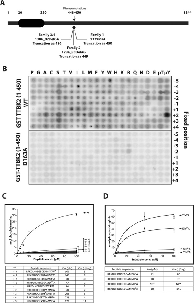
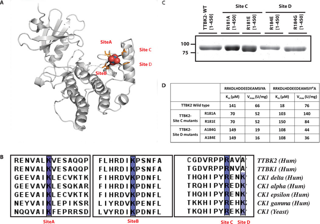
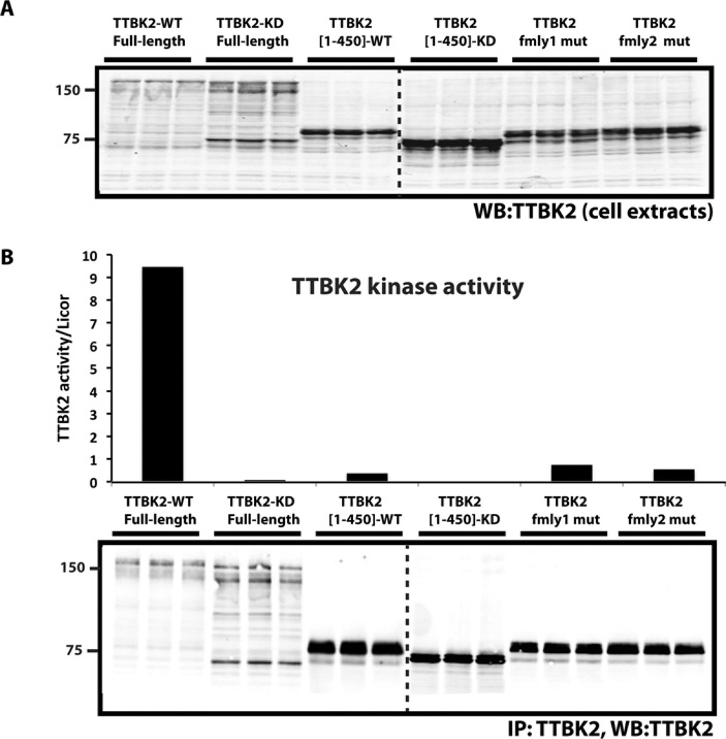
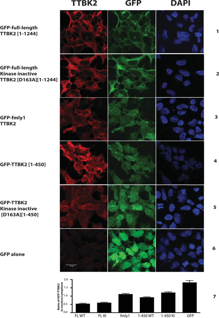
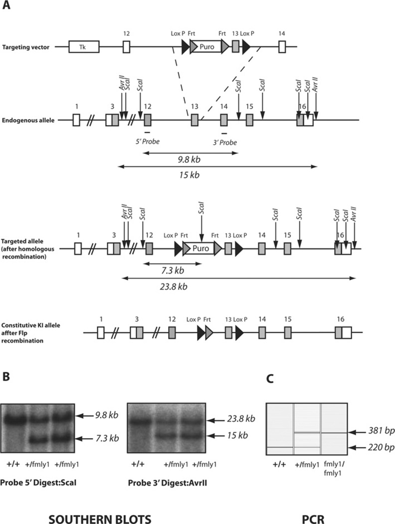
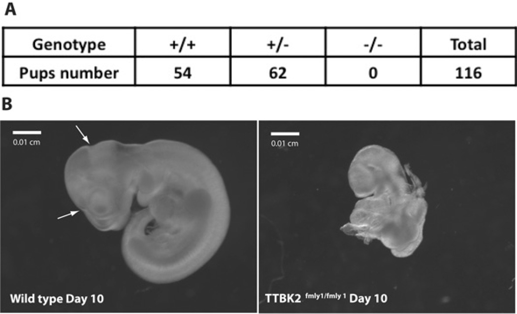
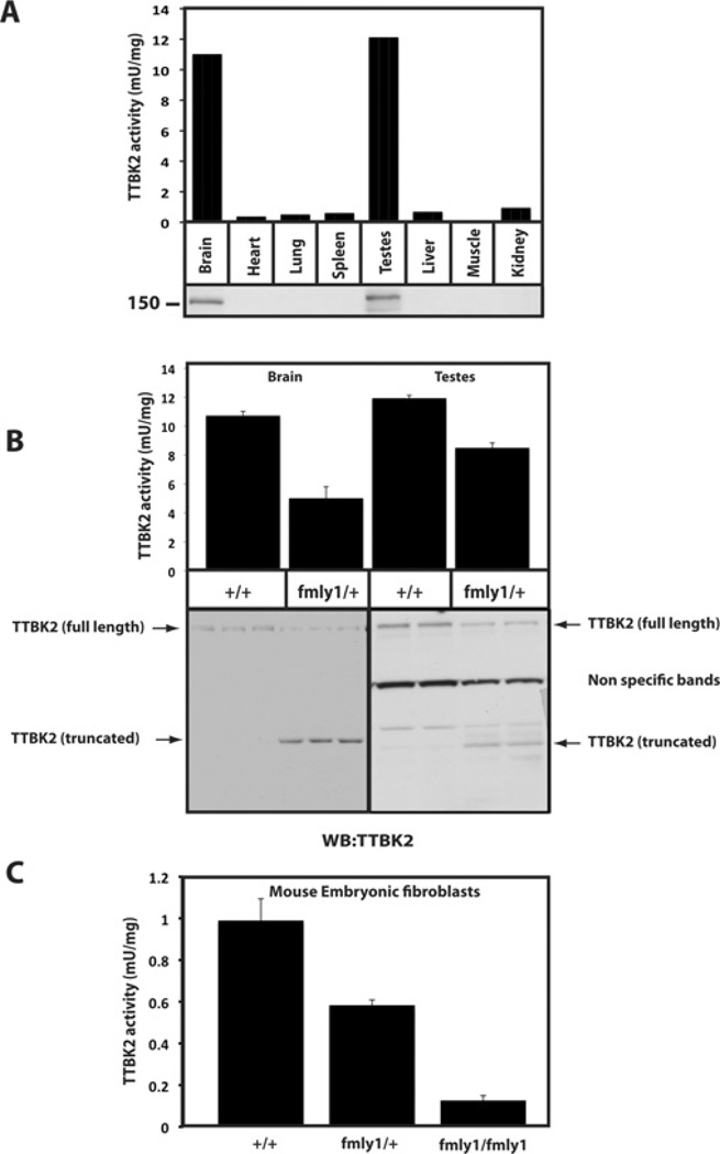
Mutations that truncate the C-terminal non-catalytic moiety of TTBK2 (tau tubulin kinase 2) cause the inherited, autosomal dominant, SCA11 (spinocerebellar ataxia type 11) movement disorder. In the present study we first assess the substrate specificity of TTBK2 and demonstrate that it has an unusual preference for a phosphotyrosine residue at the +2 position relative to the phosphorylation site. We elaborate a peptide substrate (TTBKtide, RRKDLHDDEEDEAMSIYpA) that can be employed to quantify TTBK2 kinase activity. Through modelling and mutagenesis we identify a putative phosphate-priming groove within the TTBK2 kinase domain. We demonstrate that SCA11 truncating mutations promote TTBK2 protein expression, suppress kinase activity and lead to enhanced nuclear localization. We generate an SCA11-mutation-carrying knockin mouse and show that this leads to inhibition of endogenous TTBK2 protein kinase activity. Finally, we find that, in homozygosity, the SCA11 mutation causes embryonic lethality at embryonic day 10. These findings provide the first insights into some of the intrinsic properties of TTBK2 and reveal how SCA11-causing mutations affect protein expression, catalytic activity, localization and development. We hope that these findings will be helpful for future investigation of the regulation and function of TTBK2 and its role in SCA11.


Figures

Figure 1
Figure 1. Domain structure and substrate specificity of TTBK2
(A) Schematic representation of the domain structure of TTBK2 showing the location of the three reported SCA11-causing mutations. Numbering of residues corresponds to human TTBK2. (B) Recombinant HEK-293 purified GST–TTBK2-(1–450) wild-type (WT) and catalytically inactive GST–TTBK2[D163A]-(1–450) were used to screen a positional scanning peptide library consisting of 189 biotinylated peptide libraries in individual kinase assays. Reaction products were bound to streptavidin-coated membrane and, after washing, phosphorylation was visualized by phospho-imaging. (C) Peptides with various positions of the phosphotyrosine residue from +4 to −5 relative to the phosphorylated serine residue were synthesized and analysed for their ability to phosphorylate GST–TTBK2-(1–450) WT purified from HEK-293 cells. Km and Vmax values were derived by non-linear regression analysis as described in the Materials and methods section. (D) Three TTBKtide variants with serine, threonine and tyrosine residues at the phospho-acceptor position and a TTBKtide variant with a phosphothreonine at the +2 position were synthesized, and the kinetics of their phosphorylation by GST–TTBK2-(1–450) was analysed. Km and Vmax values were derived by non-linear regression analysis. NP* denotes that the peptide was phosphorylated too poorly to undertake accurate kinetic analysis. Results in (C) and (D) are means±S.D. for three independent experiments.
Figure 2
Figure 2. Molecular basis for phosphate priming
(A) High-resolution structure of CK1δ protein showing the sulfate-binding site on the C-lobe of the kinase domain predicted to function as the phosphate-priming region [22]. Lys38 (site A), Lys130 (site B), Arg168 (site C) and Lys171 (site D) form ionic interactions with the sulfate molecule and are shown in orange. (B) Sequence alignment of the indicated species of TTBK1, TTBK2 and CK1 family enzymes showing the sequence conservation of the sulfate-binding residues. (C) Coomassie-Blue-stained gel of the GST–TTBK2-(1–450) truncated form of the indicated wild-type (WT) and mutant proteins expressed and purified from HEK-293 cells. The molecular mass in kDa is indicated on the left-hand side. (D) The indicated GST–TTBK2-(1–450) truncated forms expressed in (C) were tested for their ability to phosphorylate TTBKtide (RRKDLHDDEEDEAMSIYPA) and a variant of TTBKtide (RRKDLHDDEEDEAMSIYA) where the tyrosine at the +2 position was not phosphorylated. Km and Vmax values were derived by non-linear regression analysis.
Figure 3
Figure 3. Truncated forms of TTBK2 are less active, but are expressed at higher levels than the full-length kinase
HEK-293 cells were transiently transfected with the wild-type (WT) and indicated mutants of FLAG-tagged (A and B). (A) Cells were lysed and subjected to immunoblotting (WB) with the anti-TTBK2 antibody and the other indicated antibodies. (B) TTBK2 was immunoprecipitated (IP) from 30 µg of cell extract and subjected to a kinase-activity assay (upper panel) followed by immunoblot analysis (lower panel). TTBK2 kinase activity was quantified using 30 µM TTBKtide, and specific activity was calculated by correcting the amount of phosphate incorporation for protein levels in the immunoprecipitate using quantitative immunoblotting with the Odyssey system and is presented as c.p.m./absorbance units (c.p.m./LICOR arbitrary units). Results are means of duplicate experiments that were repeated four separate times with similar results. Dotted lines indicates that blots were on separate gels.
Figure 4
Figure 4. SCA11 disease mutations promote TTBK2 nuclear localization
HEK-293 cells stably expressing the indicated forms of GFP–TTBK2 were treated with 1 µg/ml tetracycline for 16 h to induce the expression of TTBK2 (panels 1–6). Cells were fixed with 4%PFA and stained with anti-TTBK2 antibody and with DAPI for nuclear staining. Fluorescent imaging was performed on a confocal microscope. Similar results were obtained in four independent experiments. Quantification of nuclear compared with cytoplasmic levels of TTBK2 (panel 7) was undertaken as described in the Materials and methods section. For each cell line, between 260 and 460 cells were counted over ten fields of cells; results are means±S.E.M. Scale bar, 20 µm. FL, full-length; KI, kinase inactive; WT, wild-type.
Figure 5
Figure 5. Targeting strategy used to generate TTBK2-knockin mutations
(A) Diagram describing the knockin construct, the endogenous allele containing exon 13 and the targeted allele with the puromycin (Puro) cassette removed by Flp recombinase. The grey rectangles represent TTBK2 exons. The grey and black triangles represent Frt (Flp recognition target) and LoxP sites respectively. The positions of the TTBK2 primers (P1 and P2) used for genotyping are represented as short black lines with arrowheads. KI, kinase inactive; Tk, thymidine kinase. (B) Genomic DNA purified from the targeted embryonic stem cells of the indicated genotypes was digested with either ScaI or AvrI and subjected to Southern blot analysis with the corresponding DNA probes (positions shown). In the case of the 5′ probe, the wild-type allele produces a 9.8 kb fragment and the conditional knockin allele generates a 7.3 kb fragment. Similarly, the 3′ probe detects a fragment of 23.8 kb from the wild-type allele and a 15 kb fragment from the conditional knockin allele. (C) Genomic DNA was PCR-amplified with TTBK2 primers P1 and P2. The wild-type allele (detected using P1 and P2) generates a 220 bp product and the knockin allele generates a 381 bp product.
Figure 6
Figure 6. Embryonic lethality and knockin embryo description
(A) TTBK2fmly1/+ mice were mated and the progeny genotyped as described in the Materials and methods section. The number of mice obtained is indicated for each genotype. (B) Wild-type and homozygous TTBK2fmly1/fmly1 embryos at E10 were detected at the expected Mendelian frequency. Mutant embryos are smaller and developmentally delayed, lacking prominent subdivisions of the brain (arrows on the wild-type embryo). Incomplete embryonic turning movements may result in failure to extend the body axis. A total of 27 separate E10 TTBK2fmly1/fmly1 embryos were analysed and similar phenotypes were observed.
Figure 7
Figure 7. Study of TTBK2 in wild-type and TTBK2fmly1/+ knockin mice
(A) The indicated tissue extracts were generated from wild-type mice. Extracts were immunoblotted for TTBK2 (lower panel) or TTBK2 was immunoprecipitated and subjected to a TTBK2 kinase assay employing the TTBKtide peptide substrate (upper panel). Results are means of duplicate experiments that were repeated four separate times with similar results. (B) Brain and testes lysates were generated from TTBK2+/+ and TTBK2fmly1/+ mice and subjected to immunoblot or TTBK2 kinase assay analysis, as in (A). (C) MEFs were generated from TTBK2+/+, TTBK2fmly1/+ and TTBK2fmly1/fmly1 E10 embryos as described in the Materials and methods section. TTBK2 activity was assessed following immunoprecipitation as in (A). Owing to the low levels of TTBK2 protein expressed in MEFs and high antibody background in immunoprecipitates, we were unable to detect expression of TTBK2 by immunoblot analysis. Results in (B) and (C) are means±S.D.

References

    1. Houlden H, Johnson J, Gardner-Thorpe C, Lashley T, Hernandez D, Worth P, Singleton AB, Hilton DA, Holton J, Revesz T, et al. Mutations in TTBK2, encoding a kinase implicated in tau phosphorylation, segregate with spinocerebellar ataxia type 11. Nat. Genet. 2007;39:1434–1436. - PubMed
    1. Bauer P, Stevanin G, Beetz C, Synofzik M, Schmitz-Hubsch T, Wullner U, Berthier E, Ollagnon-Roman E, Riess O, Forlani S, et al. Spinocerebellar ataxia type 11 (SCA11) is an uncommon cause of dominant ataxia among French and German kindreds. J. Neurol. Neurosurg. Psychiatry. 2010;81:1229–1232. - PubMed
    1. Houlden H. Spinocerebellar ataxia type 11. In: Pagon RA, Bird TD, Dolan CR, Stephens K, editors. GeneReviews. Seattle: University of Washington; 2008. PMID 20301723.
    1. Takahashi M, Tomizawa K, Sato K, Ohtake A, Omori A. A novel tau-tubulin kinase from bovine brain. FEBS Lett. 1995;372:59–64. - PubMed
    1. Manning G, Whyte DB, Martinez R, Hunter T, Sudarsanam S. The protein kinase complement of the human genome. Science. 2002;298:1912–1934. - PubMed

Publication types

Substances