. 2011 May 6:2:11.
doi: 10.1186/2041-9139-2-11.

Breaking evolutionary and pleiotropic constraints in mammals: On sloths, manatees and homeotic mutations

Affiliations

Breaking evolutionary and pleiotropic constraints in mammals: On sloths, manatees and homeotic mutations

Irma Varela-Lasheras et al. Evodevo. .

Erratum in

Abstract

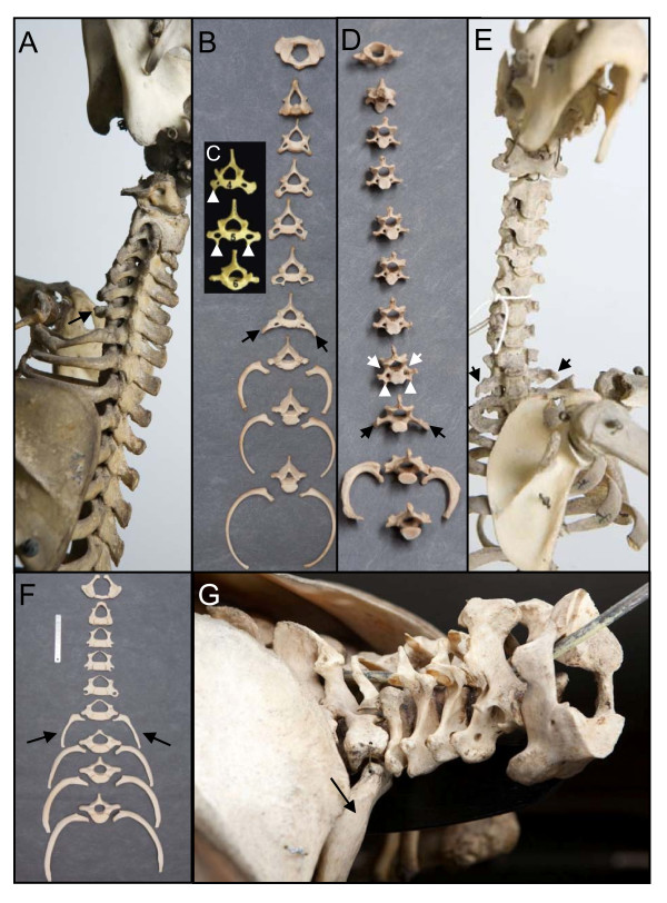
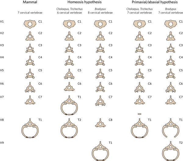
Background: Mammals as a rule have seven cervical vertebrae, except for sloths and manatees. Bateson proposed that the change in the number of cervical vertebrae in sloths is due to homeotic transformations. A recent hypothesis proposes that the number of cervical vertebrae in sloths is unchanged and that instead the derived pattern is due to abnormal primaxial/abaxial patterning.

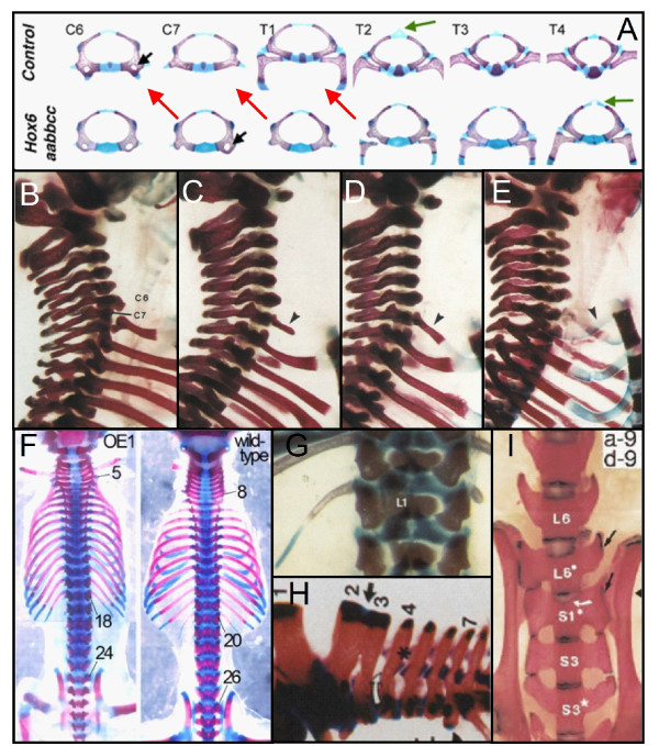
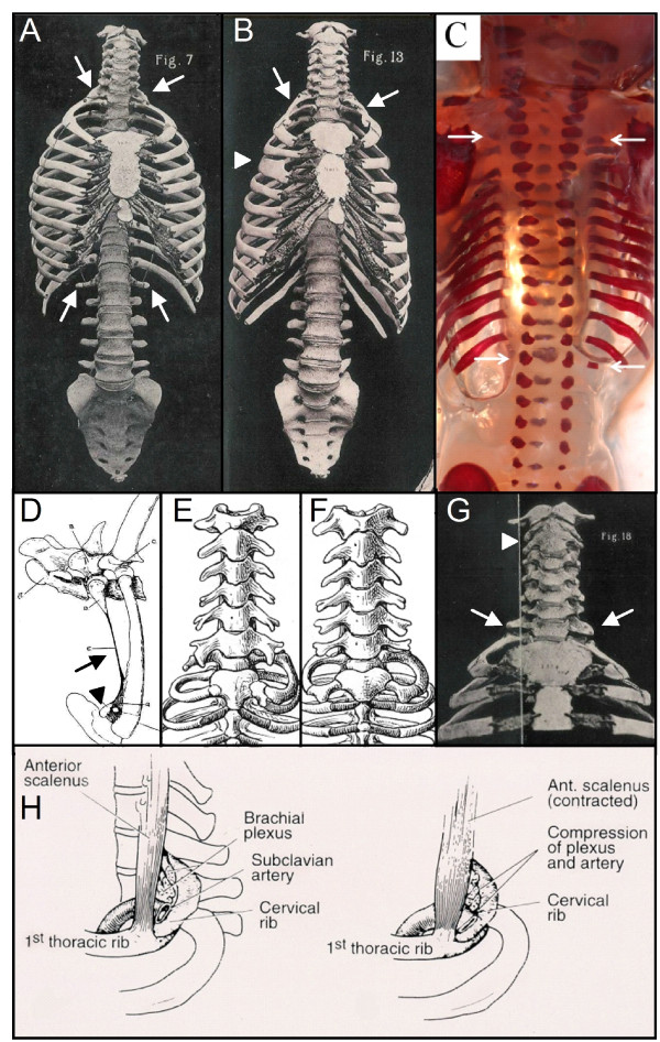
Results: We test the detailed predictions derived from both hypotheses for the skeletal patterns in sloths and manatees for both hypotheses. We find strong support for Bateson's homeosis hypothesis. The observed vertebral and rib patterns cannot be explained by changes in primaxial/abaxial patterning. Vertebral patterns in sloths and manatees are similar to those in mice and humans with abnormal numbers of cervical vertebrae: incomplete and asymmetric homeotic transformations are common and associated with skeletal abnormalities. In sloths the homeotic vertebral shift involves a large part of the vertebral column. As such, similarity is greatest with mice mutant for genes upstream of Hox.

Conclusions: We found no skeletal abnormalities in specimens of sister taxa with a normal number of cervical vertebrae. However, we always found such abnormalities in conspecifics with an abnormal number, as in many of the investigated dugongs. These findings strongly support the hypothesis that the evolutionary constraints on changes of the number of cervical vertebrae in mammals is due to deleterious pleitropic effects. We hypothesize that in sloths and manatees low metabolic and activity rates severely reduce the usual stabilizing selection, allowing the breaking of the pleiotropic constraints. This probably also applies to dugongs, although to a lesser extent.


Figures

Figure 1
Figure 1
Sloths and manatees have an abnormal number of cervical vertebrae, which can be seen from the shape of the vertebrae and the absence of ribs. A and B) Choloepus didactylus (ZMA.334 and RMNH.MAM.3274resp.) specimens with six cervical vertebrae and a seventh transitional cervico-thoracic vertebra with rudimentary rib that are fused to the vertebra (arrows). C) Anterior view of the 4th, 5th and 6th vertebrae of a Choloepus hofmanni. The fourth vertebra has an anterior tuberculum on the right side (white arrowhead) and not on the left, indicating a unilateral homeotic transformation into the 6th cervical vertebra, which is characterized by bilateral tuberculi anterior in mammals. The fifth vertebra has tuberculi anterior bilaterally (white arrowheads), indicating a complete homeotic transformation of the fifth into the sixth cervical vertebra. The 6th vertebra has a completely thoracic shape without foramina transversaria (see Figure 2) and has full ribs, indicating a homeotic transformation into the first thoracic vertebra (normally the 8th vertebra in mammals). Reproduced with permission from [56]. D) and E) Bradypus tridactylus (RMNH.MAM.10460 and ZMA.331 resp.) specimens with 8 cervical vertebrae. The 8th vertebra in D) has bilaterally foramina transversaria (white arrow) and tuberculi anterior (white arrowheads), indicating a complete homeotic transformation of the 8th vertebra into the 6th cervical vertebra. In D) and E) the ninth vertebrae have a transitional cervico-thoracic identity with no foramina transversaria and rudimentary ribs that are fused to the vertebrae (arrows). Note the asymmetric length of the ribs of the 10th vertebra. F) anterior view of five cervical vertebrae and four thoracic ones with ribs of Trichechus manatus (RMNH.MAM24221). There fifth cervical vertebrae has foramina transversaria, but no tuberculi anterior, as in the transgenic mice with loss of function of Hoxa5 [66]). The sixth vertebra has a transitional cervico-thoracic identity with no foramen transversaria, thoracic transverse processes and large cervical ribs (arrows). The seventh vertebra is the first fully thoracic vertebra with full ribs, indicating a complete homeotic transformation. G) Lateral view of Trichechus senegalensis (U. Nat coll.) with six cervical vertebrae and a completely thoracic seventh vertebra, with full ribs (arrow), indicating a complete homeotic transformation.
Figure 2
Figure 2
Schedule to show the predictions for vertebebrae shape and the presence or absence of ribs of the Homeosis hypothesis and Primaxial/abaxial hypothesis for sloths (Choloepus, Bradypus) and manatees (Trichechus). The sternal rib parts are indicated by darker coloration. Note that the sternal rib parts are usuall small in the first and second ribs of mammals.
Figure 3
Figure 3
Vertebrae in transgenic mice with homeotic mutations. A) homeotic transformations of vertebrae in Hox6 paralogous mutants (red arrows) with 8 cervical vertebrae instead of 7. Reproduced with permission from [39]. B), C), D), E) showing incomplete and complete homeotic transformations in mice with loss of function of Hoxa5. B) wild-type, C) short rudimentary rib on C7 (arrowhead), D) large rudimentary rib on C7 which is fused to the first thoracic rib (arrowhead), E) complete rib on C7 which is fused to the sternum (arrowhead). Reproduced with permission from [66]3. F) Change in the number of presacral vertebrae from 25 to 23 in transgenic mice with overexpression of Cdx1. Reproduced with permission from [97]. G) Asymmetric lumbar ribs in transgenic mice with loss of function of Hoxc8. Reproduced with permission from [138]. H) Fusion of the spinous processes of the second and third cervical vertebrae in a Hoxa4-b4 double mutant. Reproduced with permission from [138] I) Asymmetric and transitional lumbo-sacral vertebra with incomplete fusion of L6 to the sacrum (arrow). Reproduced with permission from [139].
Figure 4
Figure 4
Homeotic transformations in humans and other mammals. A) Human skeleton with rudimentary ribs on the 7th and 19th vertebrae, indicating incomplete homeotic transformations at the cervico-thoracic and thoraco-lumbar boundary (white arrows). Note the change from 24 to 23 presacral vertebrae. From [81]. B) Human skeleton with rudimentary ribs on the first thoracic vertebra (white arrows). Note the change from 24 to 25 presacral vertebrae, the abnormal shape of the fourth rib on the right (white arrowhead) and the asymmetric sternum (asymmetric transition of the manubrium to the corpus sterni). From [81]. C) Human fetal skeleton with rudimentary ribs on the 7th and unilaterally on the 19th vertebrae (white arrows). From [85]. D) Rudimentary first rib with long fibrous band (arrow) connecting to the first rib and sternum in horse (cf. Trichechus manatus with rudimentary rib and fibrous band in Figure 7F. Note that the sternal part is present, attached to the sternum (arrowhead). From [78]. E) and F), unilateral and bilateral complete rudimentary ribs in the slow lori (Nycticebus sp.). From [5]. G) Human skeleton showing rudimentary ribs on the eighth vertebra (white arrows) and a fusion of the second and third vertebra (white arrowhead). From [81]. H) The presence of a cervical rib leads to pressure on the nerves and arteries that go into the arm, especially when the anterior scalenus muscle is contracted. This may lead to Thoracic outlet syndrome. From [139].
Figure 5
Figure 5
Opposing A-P gradients of Retinoic acid (RA), Fgfs and Wnts during early organogenesis influence most processes that take place during this stage, including A-P, Medio-Lateral and Dorso-Ventral patterning of the three germ-layers of the embryo, axial lengthening, cell migration, somitogenesis and the active maintenance of bilateral symmetry of the left and right somites. Modified with permission from [139]
Figure 6
Figure 6
Transitional vertebrae in sloths and manatees showing incomplete and asymmetric transformations. A) Ventral view of a transitional thoraco-lumbar vertebra of a Bradypus tridactylus (ZMA.331) with a short rudimentary rib on the left (white arrow). B)Dorsal view of a transitional lumbo-sacral vertebra of a Choloepus didactylus (ZMA.334) with bilaterally incomplete fusion with the sacrum (arrows). C) Ventral view of a transitional sacro-coccygeal vertebra in a Bradypus tridactylus (ZMA.331) with on the right incomplete fusion with the sacrum (arrow). D) ventral view of a transitional thoraco-caudal vertebra in a Trichechus manatus (RMNH.MAM.22392),with a rudimentary rib on the right side and a transitional transverse process without rib on the left side (arrows).
Figure 7
Figure 7
Skeletal and fibrous abnormalities in sloths and manatees. A) Fusion of the dorsal spinous processes of the seventh and eighth vertebra in a Choloepus didactylus (RMNH.MAM.7203) (arrow). B) Absence of ossification in the sternum of a Choloepus didactylus (ZMA.335). C) Defective ossification of the sacrum in a Choloepus didactylus (ZMA.335) (arrow). D) Fusion of the second and third vertebra in a Trichechus senegalensis ( ZMA14042) (white arrow). E) large sternal foramen, a midline fusion defect, in a Trichechus manatus (RBINS 1.181). F) Abnormal fibrous band in a Trichechus manatus (U. Nat coll) (white arrow) that connects the rudimentary rib on the left with the sternum. Note the asymmetry of left and right ribs (white arrowheads). The position of the fibrous band suggests that the mesenchymal anlage of the rib was formed, but that there was a cell fate change from chondrification (and later ossification) to the formation of a fibrous band, see text and cf horse with rudimentary first ribs and fibrous band in Figure 4D.
Figure 8
Figure 8
Skeletal and fibrous abnormalities in individuals of Dugongs and hyracoids with an aberrant number of cervical vertebrae. A) Dendrohyrax arboreus (RMCA 22057) skeleton with full ribs on the seventh vertebra (arrows) and a sixth cervical vertebra with on the right the identity of a normal seventh vertebra and on the left of a normal sixth vertebra (with anterior tuberculum, arrowhead). B) Dugong dugon (RBINC 1.183d) with articulation facets for rudimentary ribs on the seventh cervical vertebra (arrows). Note the irregular shape of the cervical vertebral bodies, in particular of the sixth one (arrowhead). C) Dugong dugon (RMNH.MAM.27523) with a small rudimentary rib on the first caudal vertebra that is fused to the last thoracic rib (arrow). Note that the transverse process of the second caudal vertebra is fused to the rudimentary rib. D) Forefoot of a Dendrohyrax arboreus (RMCA 22057) showing four instead of five digits. Same specimen as in A) with full cervical ribs. E) Abnormally shaped scapulae of a Procavia capensis (RMCA 20098) with rudimentary cervical ribs. F) Abnormal ossification of tendons in the limb of a Dugong dugon (RBINC 1.183d) with rudimentary cervical ribs (same specimen as B). G) Dugong dugon (RMNH.MAM.27523)with fused second and third vertebrae (arrow) with a unilateral rudimentary rib (not shown).

References

    1. Colbert EU. Relationships of the Okapi. J Mammol. 1938;19:47–64. doi: 10.2307/1374281. - DOI
    1. Woolfenden GE. Postcranial morphology of the waterfowl. Bull Florida State Museum Biol Sci. 1961;6:1–129.
    1. Badlangana NL, Adams JW, Manger PR. The giraffe (Giraffa camelopardalis) cervical vertebral column: a heuristic example in understanding evolutionary processes? Zool J Linn Soc. 2009;155:736–757. doi: 10.1111/j.1096-3642.2008.00458.x. - DOI
    1. Leboucq H. Recherches sur les variations anatomiques de la première côte chez l'homme. Arch Biol. 1898;15:9–178.
    1. Schultz AH. In: Primatologia - Handbuch der Primatenkunde. Hofer H, Schultz AH, Starck D, editor. Basel/New York; 1961. Vertebral column and thorax; p. 66. Band 4, Lieferung 5.