. 2011 May 6:12:19.
doi: 10.1186/1471-2091-12-19.

Tyrosine phosphorylation of Munc18c on residue 521 abrogates binding to Syntaxin 4

Affiliations

Tyrosine phosphorylation of Munc18c on residue 521 abrogates binding to Syntaxin 4

Veronica Aran et al. BMC Biochem. .

Abstract

Background: Insulin stimulates exocytosis of GLUT4 from an intracellular store to the cell surface of fat and muscle cells. Fusion of GLUT4-containing vesicles with the plasma membrane requires the SNARE proteins Syntaxin 4, VAMP2 and the regulatory Sec1/Munc18 protein, Munc18c. Syntaxin 4 and Munc18c form a complex that is disrupted upon insulin treatment of adipocytes. Munc18c is tyrosine phosphorylated in response to insulin in these cells. Here, we directly test the hypothesis that tyrosine phosphorylation of Munc18c is responsible for the observed insulin-dependent abrogation of binding between Munc18c and Syntaxin 4.

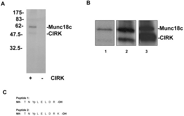
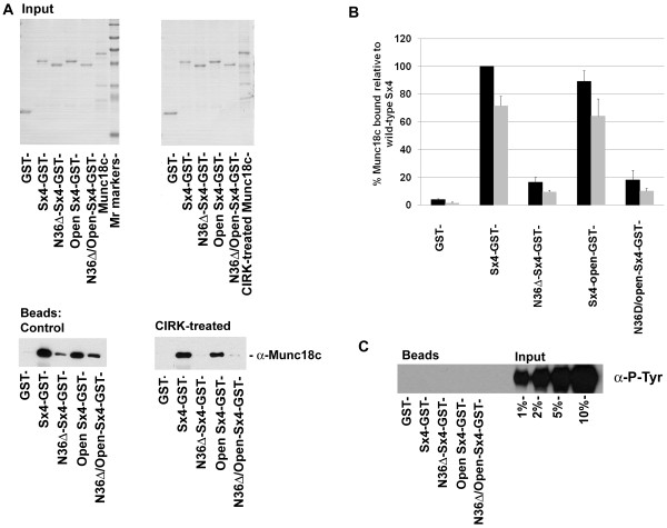
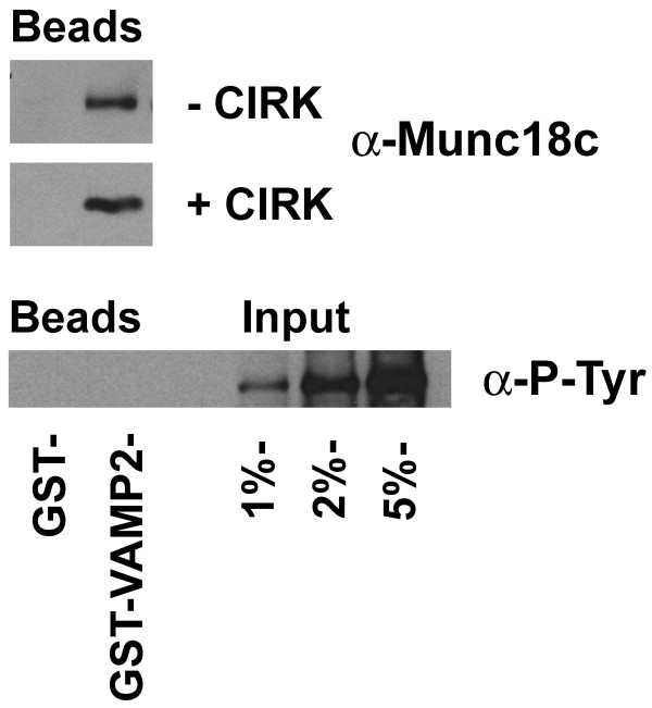
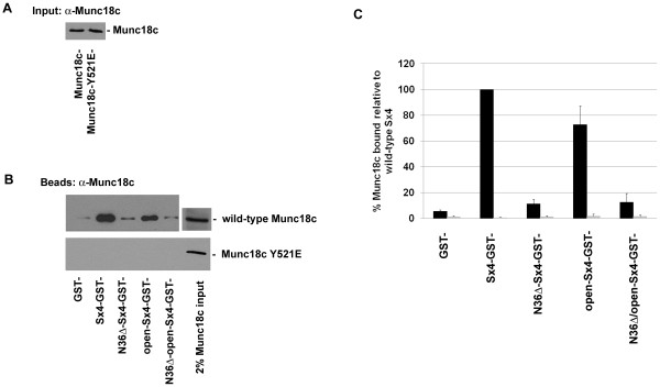
Results: We show that Munc18c is directly phosphorylated by recombinant insulin receptor tyrosine kinase in vitro. Using pull-down assays, we show that phosphorylation abrogates binding of Munc18c to both Syntaxin 4 and the v-SNARE VAMP2, as does the introduction of a phosphomimetic mutation into Munc18c (Y521E).

Conclusion: Our data indicate that insulin-stimulated tyrosine phosphorylation of Munc18c impairs the ability of Munc18c to bind its cognate SNARE proteins, and may therefore represent a regulatory step in GLUT4 traffic.


Figures

Figure 1
Figure 1
Munc18c is a substrate for CIRK in vitro. A 1 μg Munc18c was incubated with or without 370 ng CIRK in the presence of [γ-32P]-ATP. Reactions were stopped by boiling in Laemmli buffer prior to SDS-PAGE/autoradiography. Shown are data from a representative experiment. B 1 μg Munc18c was incubated with 370 ng CIRK in the presence of [γ-32P]-ATP for 30 minutes (lane 1), 120 minutes (lane 2), or 150 minutes with 1.8 μg CIRK (lane 3). Reactions were stopped as in A. Note that different exposures of the autoradiograms are shown for clarity. C Two phosphopeptides were identified by Mass Spec analysis of phosphorylated Munc18c (peptide 2 represents mis-cleavage of peptide 1).
Figure 2
Figure 2
Munc18c phosphorylated by CIRK no longer binds Sx4. A 1 μg of GST, Sx4-GST or the Sx4 mutants immobilised on glutathione-Sepharose were incubated with 1 μg of Munc18c (previously treated with or without 370 ng CIRK for 150min) in binding buffer overnight at 4°C. 6% of the samples were removed and subjected to SDS-PAGE and Coomassie stained to assess inputs (upper panels). Sepharose beads were washed prior to immunoblot analysis with anti-Munc18c (10% of eluted material; lower panels). Data shown are from representative experiments, repeated three times with similar results. B Shows quantification of the data from 3 experiments of this type. Levels of control (non-phosphorylated) Munc18c bound to the different Sx4 species is expressed as a % of the level bound to wild-type Sx4 (black bars), (mean ± SD). Data from phosphorylated Munc18c (grey bars) is also shown. Note that the level of binding of phosphorylated Munc18c to wild-type Sx4 is decreased by ~30%. Quantification of the extent of phosphorylation of Munc18c under these conditions revealed that a roughly equivalent fraction of Munc18c is phosphorylated. See text for details. The amount of Munc18c captured by the different Sx4 species is in good agreement with published data [19]. C The material eluted from the beads was also probed with anti-phosphotyrosine antibodies (40% of eluted material loaded per lane) as labelled. In this case, the indicated amounts of input were also loaded in order to confirm the presence or absence of any tyrosine phosphorylated Munc18c in the GST pull-down. Data shown are from representative experiments, repeated three times with similar results.
Figure 3
Figure 3
Munc18c phosphorylated by CIRK no longer binds VAMP2. 1 μg of GST or GST-VAMP2 immobilised on glutathione-Sepharose were incubated with 1 μg of Munc18c (which had been previously treated with or without 1.8 μg CIRK for 150min) in binding buffer overnight at 4°C. Sepharose beads were washed prior to immunoblot analysis with anti-Munc18c (upper panel) or anti-phosphotyrosine antibodies (lower panel) as labelled. In the case of the anti-phosphotyrosine immunoblot, the indicated amounts of input were also loaded to reveal the presence or absence of any tyrosine phosphorylated Munc18c in the GST pull-down. Data shown are from representative experiments, repeated three times with similar results.
Figure 4
Figure 4
Munc18c-Y521E does not bind to Sx4. 1 μg of GST, Sx4-GST or the Sx4 mutants immobilised on glutathione-Sepharose, were incubated with 1 μg of either wild-type Munc18c or Munc18c-Y521E in binding buffer overnight at 4°C. An example of the input Sx4 species is shown in Figure 2. A shows an anti-Munc18c immunoblot of wild-type Munc18c and Munc18c-Y521E recombinant proteins (3% of input in each case), indicating that both species are equally recognised by the anti-Munc18c antibody used. Sepharose beads were washed prior to immunoblot analysis with anti-Munc18c (panel B; 2% of input Munc18c or Munc18c-Y521E is also shown). Data from a representative experiment is shown, repeated three times with quantitatively similar results. We also repeated these experiments using increasing amounts of Munc18c (or the Y521E mutant) between 1 and 10 μg incubated with either 0.2 or 1.0 μg of Sx4-GST. We saw no binding of Munc18c-Y521E at any of these conditions (data not shown). Quantification of these experiments is shown graphically in panel C, black bars are wild-type Munc18c binding, grey bars are Munc18c-Y521E binding. Data presented as a % of wild-type Munc18c binding wild-type Sx4 (mean ± SD).

References

    1. Bryant NJ, Govers R, James DE. Regulated trafficking of the glucose transporter, Glut4. Nat Rev Mol Cell Biol. 2002;3:267–277. doi: 10.1038/nrm782. - DOI - PubMed
    1. Martin OJ, Lee A, McGraw TE. GLUT4 Distribution between the Plasma Membrane and the Intracellular Compartments Is Maintained by an Insulin-modulated Bipartite Dynamic Mechanism. J Biol Chem. 2006;281(1):484–490. - PubMed
    1. Hong W. SNAREs and traffic. Biochim Biophys Acta. 2005;1744(3):493–517. - PubMed
    1. Weber T, Zemelman BV, McNew JA, Westermann B, Gmachl M, Parlati F, Sollner TH, Rothman JE. SNAREpins: Minimal machinery for membrane fusion. Cell. 1998;92:759–772. doi: 10.1016/S0092-8674(00)81404-X. - DOI - PubMed
    1. Jahn R. Sec1/Munc18 proteins: mediators of membrane fusion moving to center stage. Neuron. 2000;27:201–204. doi: 10.1016/S0896-6273(00)00029-5. - DOI - PubMed

Publication types