. 2011 May 6:8:205.
doi: 10.1186/1743-422X-8-205.

Pathogenesis of Lassa fever in cynomolgus macaques

Affiliations

Pathogenesis of Lassa fever in cynomolgus macaques

Lisa E Hensley et al. Virol J. .

Abstract

Background: Lassa virus (LASV) infection causes an acute and sometimes fatal hemorrhagic disease in humans and nonhuman primates; however, little is known about the development of Lassa fever. Here, we performed a pilot study to begin to understand the progression of LASV infection in nonhuman primates.

Methods: Six cynomolgus monkeys were experimentally infected with LASV. Tissues from three animals were examined at an early- to mid-stage of disease and compared with tissues from three animals collected at terminal stages of disease.

Results: Dendritic cells were identified as a prominent target of LASV infection in a variety of tissues in all animals at day 7 while Kupffer cells, hepatocytes, adrenal cortical cells, and endothelial cells were more frequently infected with LASV in tissues of terminal animals (days 13.5-17). Meningoencephalitis and neuronal necrosis were noteworthy findings in terminal animals. Evidence of coagulopathy was noted; however, the degree of fibrin deposition in tissues was less prominent than has been reported in other viral hemorrhagic fevers.

Conclusion: The sequence of pathogenic events identified in this study begins to shed light on the development of disease processes during Lassa fever and also may provide new targets for rational prophylactic and chemotherapeutic interventions.


Figures

Figure 1
Figure 1
Hematology and coagulation values after infection of cynomolgus monkeys with Lassa virus. Development of lymphopenia (top left panel) and thromobocytopenia (top right panel). Increases in circulating levels of D-dimers (bottom left panel) and decreases in circulating levels of activated protein C (bottom right panel).
Figure 2
Figure 2
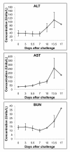
Clinical chemistry values after infection of cynomolgus monkeys with Lassa virus. Elevated levels of serum enzymes primarily at the late stages of disease (days 13-17). Top left panel, alanine aminotransferase (ALT). Top right panel, aspartate aminotransferase (AST). Bottom left panel, blood urea nitrogen (BUN).
Figure 3
Figure 3
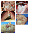
Representative gross necropsy lesions from nonhuman primates experimentally infected with Lassa virus. (A) Enlargement and moderate congestion of mesenteric lymph nodes (arrows) at day 15. (B) Accumulation of fluid in the pericardial cavity of a cynomolgus monkey 17 days after infection with Lassa virus. (C) Reticulation and discoloration of the liver 15 days after infection with Lassa virus. (D) Multifocal red foci that microscopically corresponded with inflammation on the mucosal surface of the urinary bladder at day 15. (E) Congestion/hemorrhage of the cecum occurring at day 15. The cecum is opened up and the ileum extends outward from the cecum; arrow indicates the ileocecal junction.
Figure 4
Figure 4
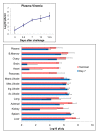
Plasma and organ titers. Mean Lassa infectivity of cynomolgus monkey plasma (top panel) and tissue homogenates (10% wt/vol) (bottom panel). LNode = lymph node.
Figure 5
Figure 5
Cytokines and chemokines. Analysis of cytokine and chemokine accumulation in serum/plasma Lassa virus-infected cynomolgus monkeys.
Figure 6
Figure 6
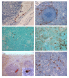
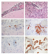
Immunohistochemistry and TUNEL staining in lymphoid tissues of cynomolgus monkeys. (A) Immunopositive endothelial cells (brown) in an axillary lymph node at day 13. (B) Immunopositive tissue macrophages and dendritic cells (brown) in spleen at day 7. (C) TUNEL-positive (brown) lymphocytes and scattered tingible body macrophages in an axillary lymph node at day 7. (D) TUNEL-positive (brown) lymphocytes and scattered tingible body macrophages in an axillary lymph node at day 17. (E) Immunopositive tissue macrophages, endothelial cells, and dendritic cells (brown) in spleen at day 13. Inset is enlargement of indicated area (arrow) showing dendritic cell. (F) Immunopositive dendritic cells (brown) in thymus at day 7. Original magnifications, ×40 (A), ×20 (C, F), ×10 (B, D, F).
Figure 7
Figure 7
PTAH staining of Lassa virus-infected cynomolgus monkey tissues. Polymerized fibrin (arrows) in the marginal zone of spleen (A), sinusoids (B) and vessels (C) in liver, and medullary vessels of the kidney (D) at day 15. Original magnifications, ×10 (D), ×20 (B), ×40 (A), ×60 (C).
Figure 8
Figure 8
Histopathology and immunohistochemistry liver and adrenal gland of Lassa virus-infected cynomolgus monkeys. Histology of liver showing inflammation at days 7 (A) and 15 (B); H&E stain. Note the progression of increased immunostaining (brown) of hepatocytes (C) and adrenal cortical cells (E) from day 7 to day 15 (D, liver; F, adrenal gland). Original magnifications, ×10 (A, B, D, F), ×20 (C, E).
Figure 9
Figure 9
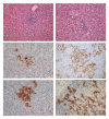
Histopathology and immunohistochemistry of nervous tissue of Lassa virus-infected cynomolgus monkeys. (A) Histology of cerebellum showing meningitis at day 15; H&E stain. (B) Histology of cerebrum showing neuronal necrosis at day 13; H&E stain. (C), Optic neuritis; note immunopositive inflammatory foci (brown) at day 15. (D), Brain stem, immunopositive endothelium (arrow) and immunopositive inflammatory foci (brown) in the neuropil peripheral to a medium-sized blood vessel at day 13. (E) Immunopositive endothelial cells (brown) in cerebrum at day 15. (F) Choroid plexus, prominent immunostaining (brown) of the cuboidal epithelial cells lining the choroid at day 15. Original magnifications, ×20 (B-F), ×40 (A)

References

    1. McCormick JB, Webb PA, Krebs JW, Johnson KM, Smith ES. A prospective study of the epidemiology and ecology of Lassa fever. J Infect Dis. 1987;155:437–444. doi: 10.1093/infdis/155.3.437. - DOI - PubMed
    1. Fisher-Hoch SP, Tomori O, Nasidi A, Perez-Oronoz GI, Fakile Y, Hutwagner L, McCormick JB. Review of cases of nosocomial Lassa fever in Nigeria: the high price of poor medical practice. Br Med J. 1995;311:857–859. - PMC - PubMed
    1. Communicable Disease Surveillance Centre. Lassa fever imported to England. Commun Dis Rep CDR Weekly. 2000;10:99. - PubMed
    1. Schmitz H, Kohler B, Laue T, Drosten C, Veldkamp PJ, Gunther S, Emmerich P, Geisen HP, Fleischer K, Beersma MF, Hoerauf A. Monitoring of clinical and laboratory data in two cases of imported Lassa fever. Microbes Infect. 2002;4:43–50. doi: 10.1016/S1286-4579(01)01508-8. - DOI - PubMed
    1. Veldkamp PJ, Schippers EF. A man with fatal Lassa fever following a stay in Sierre Leone. Ned Tijdschr Geneeskd. 2002;146:2201–2204. - PubMed

Publication types

LinkOut - more resources