. 2011 May 6:10:50.
doi: 10.1186/1476-4598-10-50.

Regulation of PBX3 expression by androgen and Let-7d in prostate cancer

Affiliations

Regulation of PBX3 expression by androgen and Let-7d in prostate cancer

Håkon Ramberg et al. Mol Cancer. .

Abstract

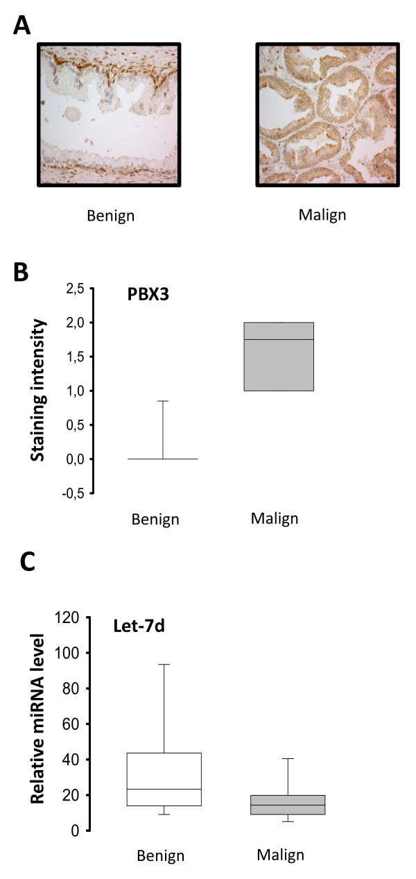
Background: The pre-leukemia transcription factor 3 (PBX) is part of the PBX family of transcription factors, which is known to regulate genes involved in differentiation of urogenital organs and steroidogenesis. This is of interest with regard to prostate cancer progression as regulation of steroidogenesis is one of the mechanisms involved in the development of castration-resistant prostate cancer. In light of this we wanted to investigate the possible involvement of androgen regulation of PBX3 expression in prostate cancer.

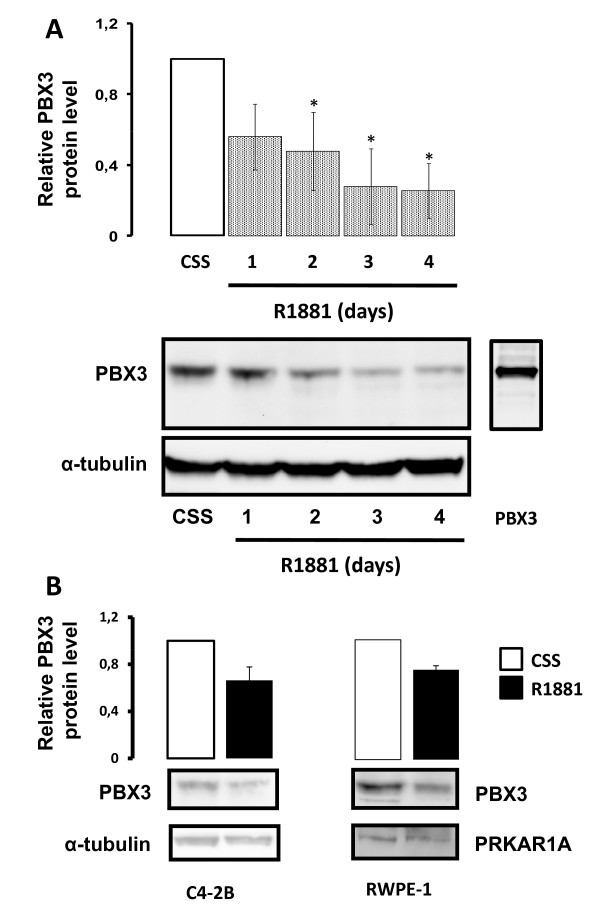
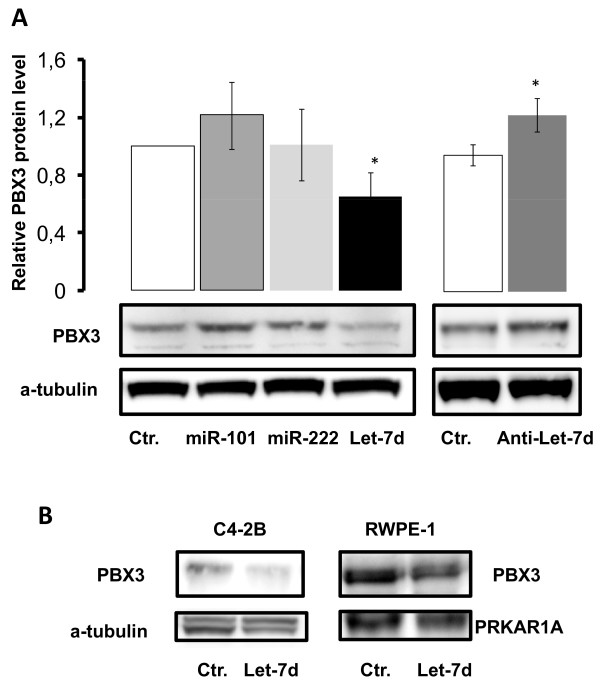
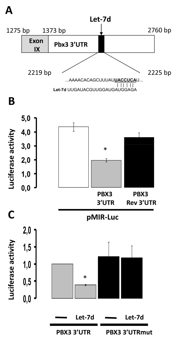
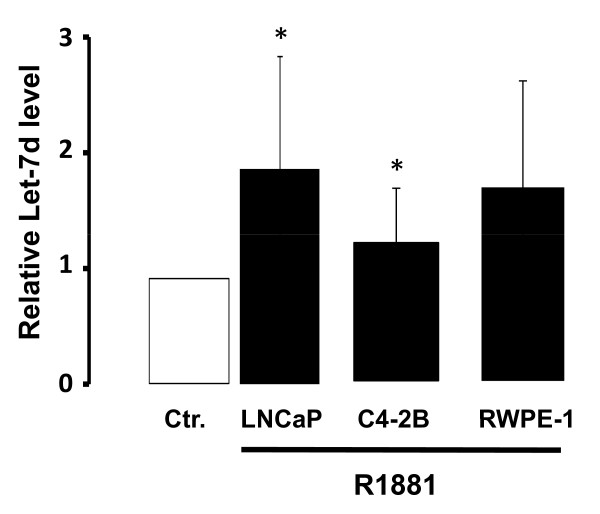
Results: In this study, we show that PBX3 is post-transcriptionally regulated by androgen in prostate cancer cells and that the effect might be independent of the androgen receptor. Furthermore, PBX3 was identified as a target of Let-7d, an androgen regulated microRNA. Let-7d was down-regulated in malignant compared to benign prostate tissue, whereas up-regulation of PBX3 expression was observed.

Conclusions: We demonstrate that PBX3 is up-regulated in prostate cancer and post- transcriptionally regulated by androgen through Let-7d.


Figures

Figure 1
Figure 1
Androgen regulation of PBX3 in prostate cancer cell lines. LNCaP cells were stimulated with 10-10M R1881 (R1881) or left untreated (CSS) for 1 - 4 days before protein were extracted. A) Representative Western blots probed with anti-human PBX3 antibody (upper panel) and anti-α-tubulin antibody as loading control (lower panel) are shown. The lane to the right shows LNCaP cells transfected with a PBX3 expression plasmid (PBX3-SPORT6). Results of densitometric analysis of the PBX3 band relative to CSS are shown in the histogram above. B) Representative Western blots of C4-2B and RWPE-1 cells stimulated with 10-10M R1881 and probed with anti-PBX3 antibody are shown in the upper panels. Anti-α-tubulin antibody (C4-2B) or anti-PRKAR1A antibody (RWPE-1) are shown as loading controls (lower panels). Data were obtained from three independent experiments and are presented in the histograms as mean ± SD (n = 3). A t-test (paired two samples of mean) was performed and a two-tailed p-value < 0, 05 is indicated with a *.
Figure 2
Figure 2
Androgen receptor independent regulation of PBX3 by R1881. A) LNCaP cells were pre-treated for 3 days with medium containing 10% CSS. Cells were then stimulated with 10-10M R1881 (R1881) and/or 10-8M bicalutamide (R1881 + Bic or Bic) or left untreated (CSS) for 4 days. Total protein extracts were analyzed by Western blotting using an anti-PBX3 antibody and anti-ERK 1/2 as loading control. Results of densitometric analysis of the PBX3 band relative to CSS are shown in the histogram. B) LNCaP cells were transfected with a small interfering RNA against androgen receptor (siRNA AR) and analyzed for PBX3 expression 48 hours post-transfection. A non-specific small interfering RNA (siRNA Ctr.) was used as control. Representative Western blots probed with anti-PBX3-, anti-AR-, anti-NKX3.1- and anti-α-tubulin antibodies are shown in the figure. The histograms depicts the results from densitometric analyses of PBX3 and AR relative to siRNA Ctr. Data were obtained from three independent experiments and are presented in the histograms as mean ± SD (n = 3). A t-test (paired two samples of mean) was performed and a two-tailed p-value < 0, 05 is indicated with a *.
Figure 3
Figure 3
MicroRNA regulation of endogenous PBX3 expression. A) LNCaP cells were transfected with 5·10-8M miR-101, miR-222, Let-7d or control mimic microRNA (Ctr.) as shown in the left panel. The right panel shows transfection of LNCaP cells with 10-7M Let-7d mimic Hairpin Inhibitor. Total protein was extracted from the cells 48 hours post-transfection. Western analysis was performed with an anti-PBX3 antibody and anti-α-tubulin as loading control. Representative Western blots are shown and the results from the densitometric analysis of three independent experiments are presented in the histograms above (mean ± SD (n = 3)). A t-test (paired two samples of mean) was performed and a two-tailed P value < 0, 05 is indicated with a *. B) Let-7d mimic transfection of C4-2B and RWPE-1 cells followed by detection of endogenous PBX3 levels by Western analysis. Anti-α-tubulin (C4-2B) and anti-PRKAR1A (RWPE-1) antibodies were used as loading controls. Representative Western blots are presented.
Figure 4
Figure 4
PBX3 is a Let-7d target gene. A) A schematic presentation of the 3'UTR sequence of PBX3 included in the reporter plasmids. The mature Let-7d sequence is shown underneath and the predicted Let-7d binding region is underlined. The underlined nucleotides are deleted in the pMIR-Luc-PBX3-3'UTR-Mut plasmid. B) LNCaP cells transiently transfected with either empty vector (pMIR-Luc) or reporter plasmids containing the 3'UTR-PBX3 in either the correct (pMIR-Luc-PBX3 3'UTR) or the inverse orientation (pMIR-Luc-PBX3 3'UTR-Rev) were harvested and reporter activity measured. C) Reporter plasmids containing the 3'UTR of PBX3 with either the wild-type binding site of Let-7d present (pMIR-Luc-Pbx3 3'UTR marked PBX3 3'UTR) or deleted (PBX3 3'UTRmut) were transfected into LNCaP cells in the absence or presence of 5·10-8 M Let-7d mimic. The cells were harvested 48 hours post-transfection. Luciferase activity adjusted for β-galactosidase activity is shown in the histograms. Data are presented as mean ± SD (n = 3) relative to LNCaP cells transfected with PBX3 3'UTR. A t-test (paired two samples of mean) was performed and a two-tailed p-value < 0, 05 is indicated with a *.
Figure 5
Figure 5
Androgen regulation of Let-7d in prostate cancer cell lines. LNCaP, C4-2 and RWPE-1 cells were stimulated with 10-10M R1881 (R1881) or left untreated (Ctr) for 24 hours. Let-7d levels were quantitated using sqRT-PCR. RNU6B microRNA was used for normalization. Data are presented as mean ± SD (n = 3) relative to the corresponding untreated cells. A t-test (paired two samples of mean) was performed and a two-tailed P value < 0, 05 is indicated with a *.
Figure 6
Figure 6
Expression of Let-7d and PBX3 in human prostate cancer specimens. A) Representative images of benign and malignant prostate tissue immunohistochemically stained with anti-human PBX3 antibody. B) TMA slides, with benign (n = 21) and malignant (n = 30) prostate tissue cores, were immunohistochemically stained with anti PBX3 antibody and scored according to staining intensity from 0 to 3. B) Levels of Let-7d in benign and malign prostate tissue samples (n = 18) from Oslo Urological University Clinic were quantified by sqRT-PCR. RNU6B microRNA was used for normalization. A Pearson's chi-squared test was performed showing a significant difference (p < 0.001). Vertical lines in each box represent the median value and Whiskers show the 95% confidence interval.

References

    1. Laurent A, Bihan R, Omilli F, Deschamps S, Pellerin I. PBX proteins: much more than Hox cofactors. Int J Dev Biol. 2008;52:9–20. doi: 10.1387/ijdb.072304al. - DOI - PubMed
    1. Schnabel CA, Selleri L, Jacobs Y, Warnke R, Cleary ML. Expression of Pbx1b during mammalian organogenesis. Mech Dev. 2001;100:131–135. doi: 10.1016/S0925-4773(00)00516-5. - DOI - PubMed
    1. Kamps MP, Murre C, Sun XH, Baltimore D. A new homeobox gene contributes the DNA binding domain of the t(1;19) translocation protein in pre-B ALL. Cell. 1990;60:547–555. doi: 10.1016/0092-8674(90)90658-2. - DOI - PubMed
    1. Nourse J, Mellentin JD, Galili N, Wilkinson J, Stanbridge E, Smith SD, Cleary ML. Chromosomal translocation t(1;19) results in synthesis of a homeobox fusion mRNA that codes for a potential chimeric transcription factor. Cell. 1990;60:535–545. doi: 10.1016/0092-8674(90)90657-Z. - DOI - PubMed
    1. Monica K, Galili N, Nourse J, Saltman D, Cleary ML. PBX2 and PBX3, new homeobox genes with extensive homology to the human proto-oncogene PBX1. Mol Cell Biol. 1991;11:6149–6157. - PMC - PubMed

Publication types

MeSH terms