. 2011 May 6:12:20.
doi: 10.1186/1471-2199-12-20.

The four Zn fingers of MBNL1 provide a flexible platform for recognition of its RNA binding elements

Affiliations

The four Zn fingers of MBNL1 provide a flexible platform for recognition of its RNA binding elements

Danielle Cass et al. BMC Mol Biol. .

Abstract

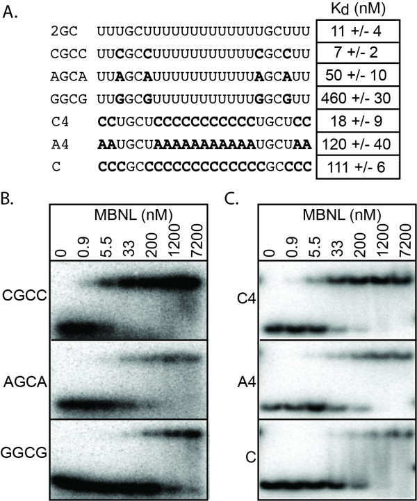
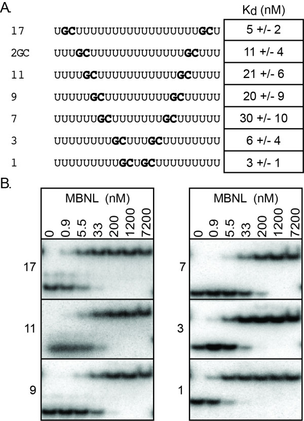
Background: Muscleblind-like 1 (MBNL1) is an alternative splicing factor containing four CCCH Zinc fingers (ZnFs). The sequestration of MBNL1 by expanded CUG and CCUG repeats is a major component in causing myotonic dystrophy. In addition to binding the structured expanded CUG and CCUG repeats; previous results suggested that MBNL1 binds single-stranded RNAs containing GC dinucleotides.

Results: We performed a systematic analysis of MBNL1 binding to single-stranded RNAs. These studies revealed that a single GC dinucleotide in poly-uridine is sufficient for MBNL1 binding and that a second GC dinucleotide confers higher affinity MBNL1 binding. However additional GC dinucleotides do not enhance RNA binding. We also showed that the RNA sequences adjacent to the GC dinucleotides play an important role in MBNL1 binding with the following preference: uridines >cytidines >adenosines >guanosines. For high affinity binding by MBNL1, the distance between the two GC dinucleotides can vary from 1 to 17 nucleotides.

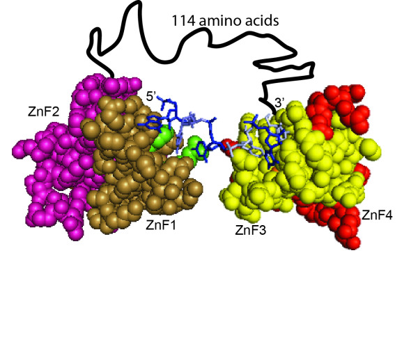
Conclusions: These results suggest that MBNL1 is highly flexible and able to adopt different conformations to recognize RNAs with varying sequence configurations. Although MBNL1 contains four ZnFs, only two ZnF - GC dinucleotide interactions are necessary for high affinity binding.


Figures

Figure 1
Figure 1
The importance of GC dinucleotides in the context of single-strand RNA for MBNL1 binding. A and E) Sequence of RNAs used in the gel shift assays to determine the specificity for the GC dinucleotides. GC dinucleotides are shown in bold with the subsequent mutations underlined. B, C and F) Representative gel shift assays. RNAs bound to MBNL1 (2-260) are labeled on the left. Concentrations of either MBNL1 or competitive RNA in each lane are at the top of the columns. D) Representative competition assay. Each lane contains 33.3 nM MBNL1, 0.02 nM radioactive 2GC RNA and increasing concentrations of competitive RNA. The identity of the competitive RNA is labeled on the left side of the gels and concentration of the competitive RNA (nM) is shown at the top of the column. On the left of each gel is the Kd based upon a competition model of binding. G) Thermal denaturation analysis from representative RNAs monitored at 260 nm. The black line is 1GC#2, red is 2GC#1, blue is 3GC#3, green is 4GC, orange is (CUG)4, and purple is 2GC. The (CUG)4 sequence is GCUGCUGUUCGCUGCUG. Refer to part A and E for the sequences of the other RNAs.
Figure 2
Figure 2
Determination of the importance of nucleotides adjacent to the GC dinucleotides for MBNL1 binding. A) RNAs used to analyze the sequence outside of the GC dinucleotides. Nucleotides that have been changed from the 2GC RNA are in bold. Next to the RNAs are the corresponding Kds, averaged over at least three independent experiments. B and C) Representative gel shifts assays. MBNL1 concentrations are at the top of each column. RNA being tested is on the left side of the gels.
Figure 3
Figure 3
Analysis of RNA spacing between GC dinucleotides on MBNL1 binding. A) RNAs used in analysis of GC spacing. Name of RNA indicates the number of nucleotides between the two GC dinucleotides. GC dinucleotides are in bold. Next to each RNA is the corresponding Kd, averaged over at least three independent experiments. B) Representative results from the gel shift assays. The RNA being tested is to the right of each gel and at the top of the column is the concentration of protein (nM) in each lane.
Figure 4
Figure 4
Model of MBNL1 binding RNA. Space-filling and stick model of MBNL1 ZnFs3/4 with CGCUGU RNA (PDB 3D2S) and MBNL1 ZnFs1/2 (PDB 3D2N). Tan = ZnF1, Magenta = ZnF2, Yellow = ZnF3, Red = ZnF4. Nucleotides making base specific contacts to ZnF3 are in white. These nucleotides come from two separate RNAs within the crystallized structure. Light blue is a cytidine from the ZnFs3/4 structure that has been modeled into the proposed cytidine binding pocket (green) of ZnF1, based upon sequence alignment with ZnFs3/4. The amino acid linker between ZnFs1/2 and ZnFs3/4 is modeled as a black line.

References

    1. Wang ET, Sandberg R, Luo S, Khrebtukova I, Zhang L, Mayr C, Kingsmore SF, Schroth GP, Burge CB. Alternative isoform regulation in human tissue transcriptomes. Nature. 2008;456:470–476. doi: 10.1038/nature07509. - DOI - PMC - PubMed
    1. Pan Q, Shai O, Lee LJ, Frey BJ, Blencowe BJ. Deep surveying of alternative splicing complexity in the human transcriptome by high-throughput sequencing. Nat Genet. 2008;40:1413–1415. doi: 10.1038/ng.259. - DOI - PubMed
    1. Sultan M, Schulz MH, Richard H, Magen A, Klingenhoff A, Scherf M, Seifert M, Borodina T, Soldatov A, Parkhomchuk D. et al.A global view of gene activity and alternative splicing by deep sequencing of the human transcriptome. Science. 2008;321:956–960. doi: 10.1126/science.1160342. - DOI - PubMed
    1. Graveley BR. Alternative splicing: increasing diversity in the proteomic world. Trends Genet. 2001;17:100–107. doi: 10.1016/S0168-9525(00)02176-4. - DOI - PubMed
    1. Hartmann B, Valcarcel J. Decrypting the genome's alternative messages. Curr Opin Cell Biol. 2009;21:377–386. doi: 10.1016/j.ceb.2009.02.006. - DOI - PubMed

Publication types

LinkOut - more resources