Review
. 2012 Jan;83(1):12-21.
doi: 10.1016/j.mvr.2011.04.005. Epub 2011 Apr 22.

Interplay between FAK, PKCδ, and p190RhoGAP in the regulation of endothelial barrier function

Affiliations
Review

Interplay between FAK, PKCδ, and p190RhoGAP in the regulation of endothelial barrier function

Katie L Grinnell et al. Microvasc Res. 2012 Jan.

Abstract

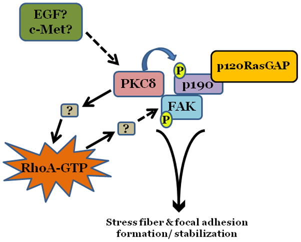
Disruption of either intercellular or extracellular junctions involved in maintaining endothelial barrier function can result in increased endothelial permeability. Increased endothelial permeability, in turn, allows for the unregulated movement of fluid and solutes out of the vasculature and into the surrounding connective tissue, contributing to a number of disease states, including stroke and pulmonary edema (Ermert et al., 1995; Lee and Slutsky, 2010; van Hinsbergh, 1997; Waller et al., 1996; Warboys et al., 2010). Thus, a better understanding of the molecular mechanisms by which endothelial cell junction integrity is controlled is necessary for development of therapies aimed at treating such conditions. In this review, we will discuss the functions of three signaling molecules known to be involved in regulation of endothelial permeability: focal adhesion kinase (FAK), protein kinase C delta (PKCδ), and p190RhoGAP (p190). We will discuss the independent functions of each protein, as well as the interplay that exists between them and the effects of such interactions on endothelial function.


Figures

Figure 1
Figure 1. PKCδ inhibition promotes endothelial barrier dysfunction
Changes in endothelial monolayer permeability were assessed in rat lung microvascular endothelial cells (LMVEC) (panel a) or endothelial cells derived from the epididymal fat pad (FPEC) (panel b) by assaying changes in resistance of endothelial monolayers grown on collagen coated gold electrodes using the electrical cell impedance system (ECIS); a drop in electrical resistance across the endothelial monolayers correlates with increased permeability. Panel a, vehicle (DMSO) or indicated concentration of rottlerin (a chemical inhibitor with specificity for PKCδ, relative to other PKC isoforms) was added to the monolayers, with arrows indicating time of addition. Panel b, endothelial monolayers containing equivalent numbers of endothelial cells were infected with indicated adenovirus. Protein overexpression was confirmed by immunoblot analyses (inset) and the effect of the overexpressed protein on monolayer permeability was determined by measuring the electrical resistance across the monolayers 24 hours post-infection. The mean±SE of the normalized electrical resistance are presented. Panels a, n=6–12; *p<0.05 vs. vehicle. Panel b, n=16; *p<0.05 vs. Ad GFP or uninfected. Panel c, pulmonary vascular permeability was measured by calculating the capillary filtration coefficients (Kf), using the Starling equation, from isolated, perfused rat lungs, which were fully recruited and in an isogravametric state. Kf was determined by measuring the lung weight gain following an increase in venous pressure divided by the change in capillary pressures and normalized to 100g wet lung mass at baseline (solid bars) and following a 45 minute exposure to vehicle (DMSO) or 50μM rottlerin (open bars). n=3–4, *p<0.05. Panels a, c, and d: Reprinted from Klinger, J.R., et al., 2007. Rottlerin causes pulmonary edema in vivo: A possible role for PKCδ. Journal of Applied Physiology, 103:2084–2094. Panel b: Reprinted from Harrington, E.O., et al., 2005. PKCδ regulates endothelial basal barrier function through modulation of RhoA GTPase activity. Experimental Cell Research, 308:407–421.
Figure 2
Figure 2. PKCδ inhibition blunts FAK activity and diminished cytoskeletal stiffness
Panel a, FAK activity was determined by measuring the level of phosphorylation at FAK Y397 by immunoblot analysis at indicated times following incubation of endothelial cells derived from the epididymal fat pad (FPEC) with 5μM rottlerin. The immunoblotted membranes were subsequently stripped and reprobed for FAK. Immunoblot signals were quantitated by densitometry and the level of FAK activity is presented as the mean±SE of the ratio of FAK Y397 phosphorylation to total FAK. Panel b, barrier function is dictated by changes in both contractile and adhesive forces, thus to measure changes in the contractile forces, cytoskeletal stiffness was assessed in lung microvascular endothelial cells (LMVEC) which were overlaid with ferrimagnetic beads, coated with the integrin receptor-specific peptide sequence (Arg-Gly-Asp; RGD), forming apical focal adhesions between the LMVEC and the ferrimagnetic beads. The beads were then twisted, using a magnet, and the resistant force was measure both before treatment (i.e., baseline) and in the same cultures 30 minutes following exposure to vehicle, 250nM Ro-31-7549 (a chemical inhibitor with specificity for PKCα, β, γ, ε), 10nM Gö6976 (a chemical inhibitor with specificity for PKCα, β, γ), or 5μM rottlerin. Data are presented as mean±SE (n=280–520 cells). *p<0.05 vs. vehicle with respective treatment. Panel a: Reprinted from Harrington, E.O., et al., 2005. PKCδ regulates endothelial basal barrier function through modulation of RhoA GTPase activity. Experimental Cell Research, 308:407–421. Panel b: Reprinted from Klinger, J.R., et al., 2007. Rottlerin causes pulmonary edema in vivo: A possible role for PKCδ. Journal of Applied Physiology, 103:2084–2094.
Figure 3
Figure 3. PKCδ activity inversely affects p190 activity
Confluent lung microvascular endothelial cells (LMVEC) were treated with vehicle (DMSO) or 10μM rottlerin for 30 minutes (panel a) or infected with adenoviral vectors encoding GFP or wild-type PKCδ cDNA (panel b). Cells were harvested and p190 activity determined as the level of p190 bound to GST-fused constitutively actvated RhoA. The level of active p190 relative to total p190 was determined by densitometry. In panel b, GFP and PKCδ overexpression was confirmed in the transfected endothelial cells by immunoblot analysis. Data are presented as the mean±SE. Panel a, n=4; *p<0.05 vs. vehicle. Panel b, n=7, #p<0.05 vs. GFP. Panels a and b: Reprinted from Fordjour, A.K. and Harrington, E.O. 2009. PKCδ influences p190 phosphorylation and activity: Events independent of PKCδ-mediated regulation of endothelial cell stress fiber and focal adhesion formation and barrier function. Biochimica et Biophysica Acta, 1790:1179–1190.
Figure 4
Figure 4. Model of potential cross talk between PKCδ, p190, and FAK
Under basal conditions, PKCδ functions to maintain endothelial barrier function through maintenance of a static level of RhoA activation and stimulation of FAK autophosphorylation. This allows for focal adhesion stabilization and organization of actin stress fibers. PKCδ-mediated activation of RhoA appears to occur independently of its effects on p190 phosphorylation.

Similar articles

Cited by

References

    1. Adamson P, Etienne S, Couraud PO, Calder V, Greenwood J. Lymphocyte migration through brain endothelial cell monolayers involves signaling through endothelial ICAM-1 via a rho-dependent pathway. Journal of Immunology. 1999;162(5):2964–2973. - PubMed
    1. Alghisi GC, Ponsonnet L, Rüegg C. The integrin antagonist cilengitide activates αVβ3, disrupts VE-cadherin localization at cell junctions and enhances permeability in endothelial cells. PLoS One. 2009;4:e4449. - PMC - PubMed
    1. Almeida EA, et al. Matrix survival signaling: from fibronectin via focal adhesion kinase to c-Jun NH(2)-terminal kinase. Journal of Cell Biology. 2000;149:741–754. - PMC - PubMed
    1. Anfuso CD, et al. Endothelial cell-pericyte cocultures induce PLA2 protein expression through activation of PKCα and the MAPK/ERK cascade. Journal of Lipid Research. 2007;48(4):782–793. - PubMed
    1. Arthur WT, Burridge K. RhoA inactivation by p190RhoGAP regulates cell spreading and migration by promoting membrane protrusion and polarity. Molecular Biology of the Cell. 2001;12(9):2711–2720. - PMC - PubMed

Publication types

MeSH terms

Substances

LinkOut - more resources