. 2011 Jul;91(1):1-10.
doi: 10.1016/j.antiviral.2011.04.009. Epub 2011 Apr 23.

Interference of ribosomal frameshifting by antisense peptide nucleic acids suppresses SARS coronavirus replication

Affiliations

Interference of ribosomal frameshifting by antisense peptide nucleic acids suppresses SARS coronavirus replication

Dae-Gyun Ahn et al. Antiviral Res. 2011 Jul.

Abstract

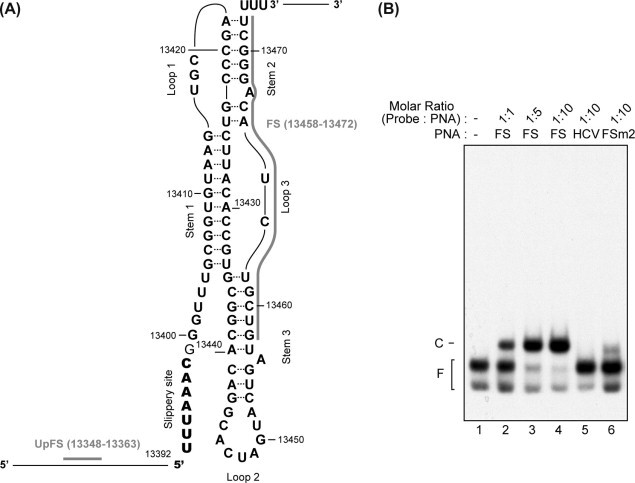
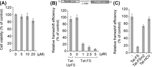
The programmed -1 ribosomal frameshifting (-1 PRF) utilized by eukaryotic RNA viruses plays a crucial role for the controlled, limited synthesis of viral RNA replicase polyproteins required for genome replication. The viral RNA replicase polyproteins of severe acute respiratory syndrome coronavirus (SARS-CoV) are encoded by the two overlapping open reading frames 1a and 1b, which are connected by a -1 PRF signal. We evaluated the antiviral effects of antisense peptide nucleic acids (PNAs) targeting a highly conserved RNA sequence on the - PRF signal. The ribosomal frameshifting was inhibited by the PNA, which bound sequence-specifically a pseudoknot structure in the -1 PRF signal, in cell lines as assessed using a dual luciferase-based reporter plasmid containing the -1 PRF signal. Treatment of cells, which were transfected with a SARS-CoV-replicon expressing firefly luciferase, with the PNA fused to a cell-penetrating peptide (CPP) resulted in suppression of the replication of the SARS-CoV replicon, with a 50% inhibitory concentration of 4.4μM. There was no induction of type I interferon responses by PNA treatment, suggesting that the effect of PNA is not due to innate immune responses. Our results demonstrate that -1 PRF, critical for SARS-CoV viral replication, can be inhibited by CPP-PNA, providing an effective antisense strategy for blocking -1 PRF signals.


Figures

Fig. 1
Fig. 1
Secondary structure of programmed −1 ribosomal frameshift signal and specific binding of FS PNA to its target site. (A) PNA target sites are depicted in the secondary structure of −1 PRF signal by thick lines. PNAs were designed to be complementary to the target sites. (B) The 5′-end 32P-labeled RNA probe (10 fmol) was incubated with FS PNA, FSm2 PNA, or HCV PNA in various probe–PNA molar ratios. The mixtures were resolved on a 5% non-denaturing polyacrylamide gel. Free probes (F) and probe–PNA complexes (C) are indicated.
Fig. 2
Fig. 2
Inhibition of −1 PRF by CPP-PNA in a cell-based assay. (A) The cytotoxicity of FS PNA in Vero cells was evaluated by the MTT reduction method. Data are expressed as percentages of cell viability in the untreated cells. (B) Vero cells treated with various concentrations of Tat-UpFS or Tat-FS PNAs were transfected with a reporter plasmid harboring the −1 PRF signal (pJD502; upper panel) for measuring frameshifting efficiency, or its derivative, pJD464, which was used as a zero-frame control plasmid. Reporter plasmids were transfected into the cells using DMRIE-C transfection reagent. At 24 h post-transfection, cells were harvested and the ribosomal frameshifting assay was performed. Data are expressed as percentages of normalized firefly luciferase activity in mock-treated cells. (C) Sequence-specific inhibition of frameshifting of PNAs were assessed as in (B) with 5 μM of each indicated PNA. Reporter plasmids were transfected into the cells using Fugene 6. All data in (A–C) represent means ± SD of triplicate measurements from three independent experiments.
Fig. 3
Fig. 3
A SARS-CoV replicon expressing a luciferase reporter. (A) The Feo gene fused or non-fused to TRS9 was inserted into pBAC-SARS-CoV-REP (REP) to construct pSARS-CoV-REP-Feo (Feo) or pSARS-CoV-REP-ΔTRSFeo (ΔTRS), respectively. The pSARS-CoV-REP-Feo-MluIrev (MluIrev), which is defective in synthesis of functional replicase proteins, was used as a negative control plasmid. TRSs are indicated by a black box and leader sequences by a box with deviant crease lines. Black arrows indicate primers used for detection of N gene-specific sg-mRNAs and gray arrows for detection of sg-mRNAs containing the Feo gene. (B and C) BHK-21 cells were co-transfected with replicon plasmids and pRL-TK used for normalization of transfection efficiency, by electroporation. At 30 h post-transfection, cells were harvested and analyzed for sg-mRNA level, luciferase activity, and intracellular SARS-CoV nucleocapsid N protein level. (B) The N gene-specific sg-mRNA level was quantified by real-time qRT-PCR using a TaqMan probe. Subgenomic RNA copy numbers per μg total RNA are shown. ND, not detected. (C) Firefly luciferase activity from the replicon plasmid was normalized to Renilla luciferase activity from the pRL-TK plasmid. Normalized luciferase activity of cells transfected with pSARS-CoV-REP was defined as 100. Endogenous sg-mRNAs containing Feo gene was amplified by RT-PCR and resulting PCR products were resolved by agarose gel electrophoresis (low panel). (D) BHK-21 or HEK293 cells were left untransfected (Mock) or transfected with the plasmid indicated above the blots. N protein and α-tubulin were detected by Western blot analysis. (E) Kinetics of SARS-CoV replicon replication in transiently transfected cells. BHK-21 cells were transfected with pSARS-REP-Feo (●) or pSARS-REP-ΔTRSFeo (▴) by electroporation. Cells were harvested at each given time point and store at −80 °C until analysis. Luciferase activity was measured with the same amount of cell lysate. Data from one representative experiment from two independent experiments with similar results are shown.
Fig. 4
Fig. 4
Suppression of SARS-CoV replication by Tat-peptide-conjugated FS PNA in SARS-CoV replicon-replicating cells. (A) HEK293 cells, co-transfected with pSARS-REP-Feo and pRL-TK, were treated with each indicated PNA (10 μM) in serum-free DMEM for 3 h at 6 h post-transfection. Tat-conjugated J3U2 PNA (Tat-J3U2) targeting the 3′-UTR of JEV genome was used as a negative control. IFN-β (250 IU/ml) and IFN-β inducer poly(I:C) (0.8 μg/ml) were used as positive controls. Poly(I:C) was transfected into the cells using lipofectamin RNAiMAX agent. After 30 h incubation in complete medium, cells were harvested and luciferase activity was measured. (B) The IC50 value for the inhibition of SARS-CoV replication by Tat-FS PNA was determined at various PNA concentrations. (C) HEK293 cells were co-transfected with IFNβ-pGL3 luciferase reporter plasmid and pRL-TK used for normalization of transfection efficiency, prior to mock-treatment or treatment with 10 μM of Tat-FS or Tat-FSm2 PNA. Poly(I:C) or HCV 3′-UTR at a final concentration of 2 μg/ml or expression of an active form of IRF3, IRF3(5D) was used as a positive control for stimulation of IFN-β promoter activity. The normalized firefly luciferase activity of the mock-treated cells is considered one unit and the increase in luciferase activity is shown as fold induction. (D) HEK293 cells treated with PNAs or indicated RNAs as in (C) were analyzed for IFN-β and ISG56 mRNA abundance by real-time qRT-PCR. Fold increase in mRNA abundance is shown. (E) The real-time PCR products representing the abundance of the indicated mRNAs were visualized by agarose gel electrophoresis and ethidium bromide staining. Detection of GAPDH mRNA served as control. Results are from a representative experiment of n = 3 that gave similar results. (A–D) Data represent means ± SD of triplicate measurements from three independent experiments.

Similar articles

Cited by

References

    1. Almazan F., Dediego M.L., Galan C., Escors D., Alvarez E., Ortego J., Sola I., Zuniga S., Alonso S., Moreno J.L., Nogales A., Capiscol C., Enjuanes L. Construction of a severe acute respiratory syndrome coronavirus infectious cDNA clone and a replicon to study coronavirus RNA synthesis. J. Virol. 2006;80:10900–10906. - PMC - PubMed
    1. Baranov P.V., Gesteland R.F., Atkins J.F. Recoding: translational bifurcations in gene expression. Gene. 2002;286:187–201. - PubMed
    1. Baranov P.V., Henderson C.M., Anderson C.B., Gesteland R.F., Atkins J.F., Howard M.T. Programmed ribosomal frameshifting in decoding the SARS-CoV genome. Virology. 2005;332:498–510. - PMC - PubMed
    1. Brierley I., Digard P., Inglis S.C. Characterization of an efficient coronavirus ribosomal frameshifting signal: requirement for an RNA pseudoknot. Cell. 1989;57:537–547. - PMC - PubMed
    1. Cinatl J., Jr., Michaelis M., Hoever G., Preiser W., Doerr H.W. Development of antiviral therapy for severe acute respiratory syndrome. Antiviral Res. 2005;66:81–97. - PMC - PubMed

Publication types

MeSH terms