

Eyes wide open: a critical review of sphere-formation as an assay for stem cells
- PMID: 21549325
- PMCID: PMC3633588
- DOI: 10.1016/j.stem.2011.04.007
Eyes wide open: a critical review of sphere-formation as an assay for stem cells
Abstract
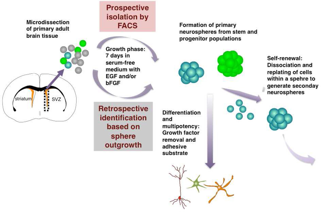
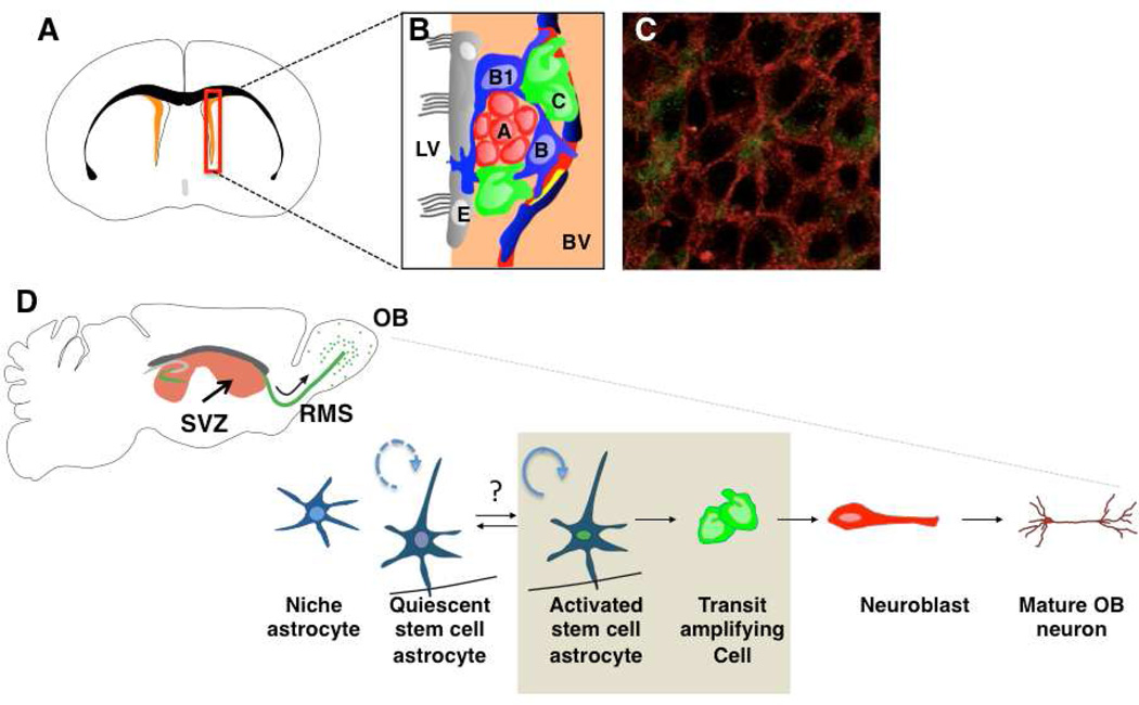
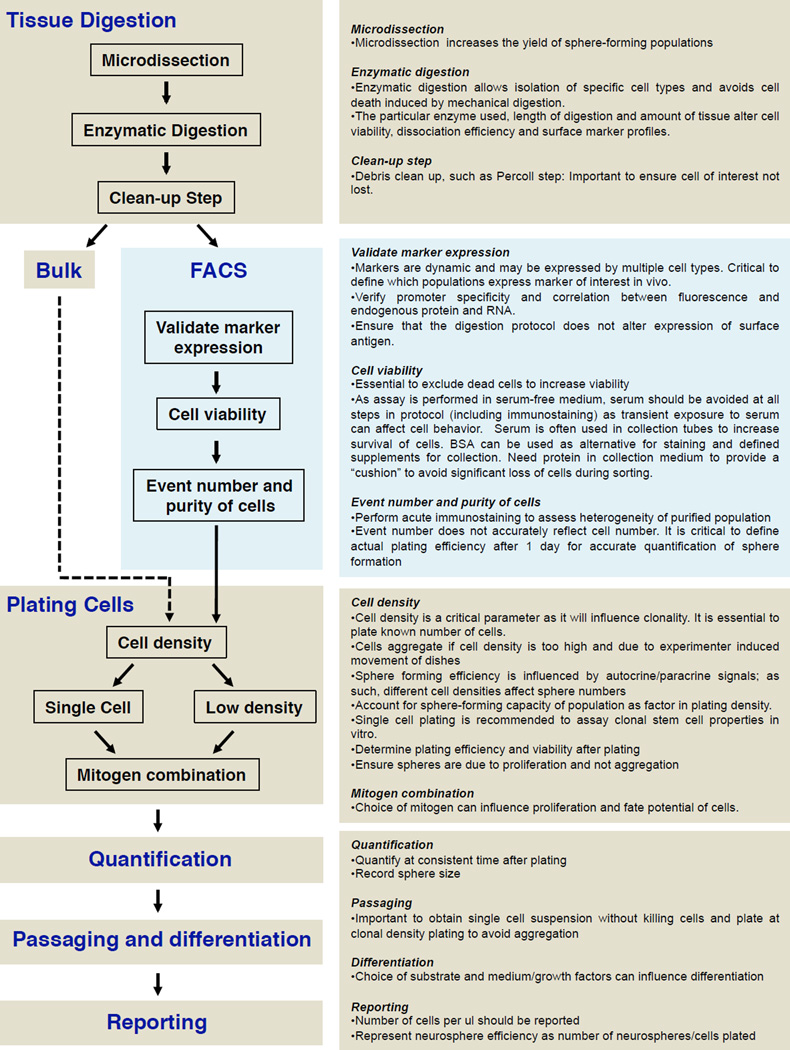
Sphere-forming assays have been widely used to retrospectively identify stem cells based on their reported capacity to evaluate self-renewal and differentiation at the single-cell level in vitro. The discovery of markers that allow the prospective isolation of stem cells and their progeny from their in vivo niche allows the functional properties of purified populations to be defined. We provide a historical perspective of the evolution of the neurosphere assay and highlight limitations in the use of sphere-forming assays in the context of neurospheres. We discuss theoretical and technical considerations of experimental design and interpretation that surround the use of this assay with any tissue.
Copyright © 2011 Elsevier Inc. All rights reserved.
Figures
References
-
- Altman J. Autoradiographic and histological studies of postnatal neurogenesis. IV. Cell proliferation and migration in the anterior forebrain, with special reference to persisting neurogenesis in the olfactory bulb. J. Comp. Neurol. 1969;137:433–457. - PubMed
-
- Andreu-Agulló C, Morante-Redolat JM, Delgado AC, Fariñas I. Vascular niche factor PEDF modulates Notch-dependent stemness in the adult subependymal zone. Nat. Neurosci. 2009;12:1514–1523. - PubMed
-
- Beckervordersandforth R, Tripathi P, Ninkovic J, Bayam E, Lepier A, Stempfhuber B, Kirchhoff F, Hirrlinger J, Haslinger A, Lie DC, Beckers J, Yoder B, Irmler M, Götz M. In vivo fate mapping and expression analysis reveals molecular hallmarks of prospectively isolated adult neural stem cells. Cell Stem Cell. 2010;7:744–758. - PubMed
-
- Barraud P, Thompson L, Kirik D, Björklund A, Parmar M. Isolation and characterization of neural precursor cells from the Sox1-GFP reporter mouse. Eur. J. Neurosci. 2002;22:1555–1569. - PubMed
Publication types
MeSH terms
Grants and funding
LinkOut - more resources
Full Text Sources
Other Literature Sources
Medical
