. 2011 May 6;8(5):552-65.
doi: 10.1016/j.stem.2011.02.021.

Nerve-derived sonic hedgehog defines a niche for hair follicle stem cells capable of becoming epidermal stem cells

Affiliations

Nerve-derived sonic hedgehog defines a niche for hair follicle stem cells capable of becoming epidermal stem cells

Isaac Brownell et al. Cell Stem Cell. .

Abstract

In adult skin, stem cells in the hair follicle bulge cyclically regenerate the follicle, whereas a distinct stem cell population maintains the epidermis. The degree to which all bulge cells have equal regenerative potential is not known. We found that Sonic hedgehog (Shh) from neurons signals to a population of cells in the telogen bulge marked by the Hedgehog response gene Gli1. Gli1-expressing bulge cells function as multipotent stem cells in their native environment and repeatedly regenerate the anagen follicle. Shh-responding perineural bulge cells incorporate into healing skin wounds where, notably, they can change their lineage into epidermal stem cells. The perineural niche (including Shh) is dispensable for follicle contributions to acute wound healing and skin homeostasis, but is necessary to maintain bulge cells capable of becoming epidermal stem cells. Thus, nerves cultivate a microenvironment where Shh creates a molecularly and phenotypically distinct population of hair follicle stem cells.


Figures

Figure 1
Figure 1
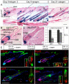
Hh-responding cells are localized in molecularly distinct subdomains of the telogen hair follicle. Also see Figures S1, S2. (A) X-gal staining in adult Gli1LacZ/+ skin at day 0, 9, and 21 after depilation of telogen hair to induce regeneration of the anagen follicle. b; bulge, Scale bars=100μm. (B, C) X-gal staining of Gli1LacZ/+ and Ptc1LacZ/+ telogen follicles showing Hh-response genes in the upper bulge, lower bulge, HG, and DP. Scale bars=100μm. (D) Relative expression levels of Gli1 and Ptc1 mRNA assessed by RT-qPCR in wild type telogen skin treated with Hh-neutralizing antibody. Error bars=SEM. club; club hair. (E) Immunostaining of Gli1LacZ and the progenitor cell markers indicated. Blue; DAPI. *Non-specific staining. Scale bar=100μm.
Figure 2
Figure 2
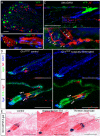
Shh from sensory nerves signals to K15(−) hair follicle epithelium in the upper telogen bulge. Also see Figures S3, S4. (A-C) Immunostaining in adult Shh-GIFM mice after TM induction. A) DRG. Scale bar=100μm. B) Axial and longitudinal sections through cutaneous nerves. NF; neurofilament. Scale bars=10μm. C) Hair follicle. Inset; magnification of boxed area, arrowheads; YFP(+) nerve endings on hair follicle. Scale bar=100μm. (D) Immunostaining of Gli1LacZ/+ skin showing changes in the telogen bulge after denervation. Arrowheads; normally K15(−) upper bulge. Scale bar=100μm. (E) X-gal staining of Gli1-GIFM telogen follicles. Upper bugle cells are not labeled in skin that is denervated then induced with TM (denervate then TM). Labeled upper bulge cells persist in skin denervated after TM induction (TM then denervate). Scale bars=100μm.
Figure 3
Figure 3
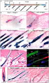
Gli1(+) cells in the telogen hair follicle regenerate the anagen follicle and self renew for normal lifespan of the animal. Also see Figure S5. (A) Scheme of experiment with TM induction of Gli1-GIFM mice in telogen phase of hair cycle prior to depilation. (B-E) X-gal staining of Gli1-GIFM skin throughout hair cycle showing expansion of initially labeled cells during anagen regrowth. Scale bars=100μm. (F, H) X-gal staining of fate-mapped cells in Gli1-GIFM skin 1 year after labeling during telogen. Labeled cells retain the ability to regenerate the anagen follicle. Arrowhead; labeling in bulge region, outline; DP. Scale bars=100μm. (G) Immunostaining anagen follicle in same mice given EdU, showing proliferating GIFM labeled cells. Arrowheads; Edu(+) ßgal(+) cells in ORS. Scale bar=100μm. (I) X-gal staining of control and denervated Gli1-GIFM skin induced with TM during telogen and collected 14 days after anagen onset. Scale bars=100μm.
Figure 4
Figure 4
Gli1(+) cells in the telogen hair follicle are multipotent. (A) Whole mount X-gal staining in anagen follicles isolated from Gli1-GIFM skin induced at clonal frequency during telogen. Examples of labeling in multiple epithelial lineages, a single lineage (HS), bulge only, and in the DP. Arrowhead; labeling in bulge region. Scale bar=100μm. (B) Frequency of anagen follicles with labeling after clonal Gli1-GIFM during telogen. follicle clones; any epithelial lineage. (C) Frequency of staining in epithelial lineage compartments among labeled follicles. single; restricted to one lineage compartment, multiple (multi); at least two lineages.
Figure 5
Figure 5
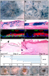
Wound healing converts Gli1(+) hair follicle cells into epidermal stem cells. Also see Figure S6. (A-B) X-gal staining of whole mount healed wounds and sections of regenerated epidermis in Gli1-GIFM mice induced during telogen, 2 weeks and 12 months after full-thickness skin wounding. Dashed line; wound area, dotted line; epidermal basement membrane, epi; regenerated epidermis. A, B: scale bars=1mm, A’, B’: scale bars=100μm. (C-D) Immunostaining of regenerated epidermis in Gli1-GIFM mice 12 months after wounding. dotted line; epidermal basement membrane. scale bars=10μm. (E) X-gal staining in Gli1LacZ/+ skin, 3 days and 10 days after wounding. Arrow; edge of wound, dashed line; regenerating epidermal tongue. Scale bars=100μm. (F) Experimental scheme to test when Gli1-GIFM cells exit follicle into healing wound. (G-K) Whole mount X-gal staining 2 weeks after wounding in Gli1-GIFM mice induced with TM on different days relative to wounding. Scale bar=1mm.
Figure 6
Figure 6
Perineural niche is required to maintain bulge cells that convert to epidermal stem cells after wounding. (A) Whole mount X-gal staining 2 weeks and 2 months after wounding in Gli1-GIFM skin with TM induction during telogen and denervation prior to or following TM. Dashed line; wound area. Scale bars=1mm. (B) Percent of regenerated epidermis with labeled cells 2 months after wounding. Error bars=SEM.
Figure 7
Figure 7
Schematic summarizing expression domains of stem cell markers and the functionally distinct perineural subdomain in the telogen hair follicle. Expression of Gli1LacZ and other markers define molecularly distinct zones in the telogen follicle, including regionalization of the bulge into the upper, middle, and lower bulge. Gli1(+) cells in the upper bulge (upper Gli1) receive Shh signaling from follicle-associated nerve endings and are functionally distinct from the cells in the middle bulge, lower bulge and HG in their ability to become epidermal stem cells during wound healing.

Comment in

Similar articles

Cited by

References

    1. Adolphe C, Narang M, Ellis T, Wicking C, Kaur P, Wainwright B. An in vivo comparative study of sonic, desert and Indian hedgehog reveals that hedgehog pathway activity regulates epidermal stem cell homeostasis. Development. 2004;131:5009–5019. - PubMed
    1. Ahn S, Joyner AL. Dynamic changes in the response of cells to positive hedgehog signaling during mouse limb patterning. Cell. 2004;118:505–516. - PubMed
    1. Ahn S, Joyner AL. In vivo analysis of quiescent adult neural stem cells responding to Sonic hedgehog. Nature. 2005;437:894–897. - PubMed
    1. Bai CB, Auerbach W, Lee JS, Stephen D, Joyner AL. Gli2, but not Gli1, is required for initial Shh signaling and ectopic activation of the Shh pathway. Development. 2002;129:4753–4761. - PubMed
    1. Bai CB, Joyner AL. Gli1 can rescue the in vivo function of Gli2. Development. 2001;128:5161–5172. - PubMed

Publication types

MeSH terms

Associated data