Case Reports
. 2011 May 13;88(5):650-6.
doi: 10.1016/j.ajhg.2011.04.010. Epub 2011 May 5.

Exome sequencing and functional analysis identifies BANF1 mutation as the cause of a hereditary progeroid syndrome

Affiliations
Case Reports

Exome sequencing and functional analysis identifies BANF1 mutation as the cause of a hereditary progeroid syndrome

Xose S Puente et al. Am J Hum Genet. .

Abstract

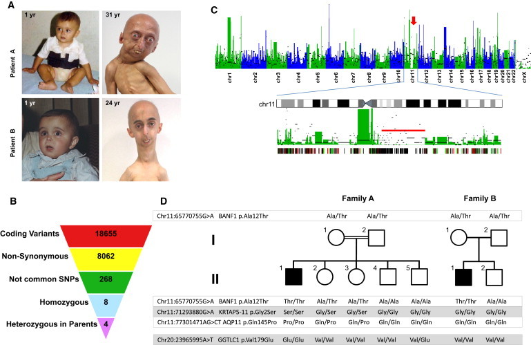
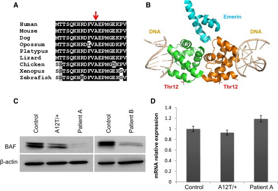
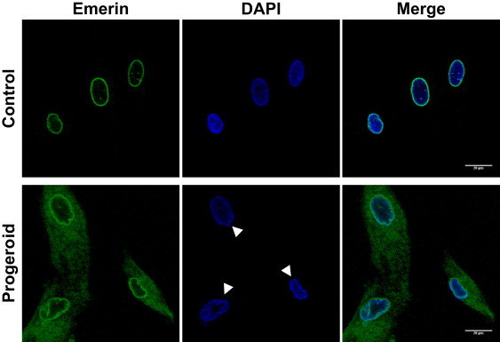
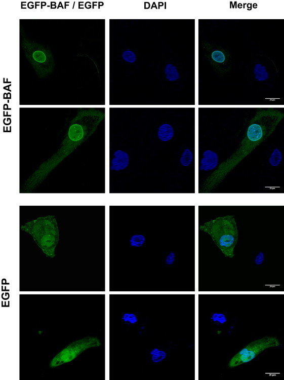
Accelerated aging syndromes represent a valuable source of information about the molecular mechanisms involved in normal aging. Here, we describe a progeroid syndrome that partially phenocopies Hutchinson-Gilford progeria syndrome (HGPS) but also exhibits distinctive features, including the absence of cardiovascular deficiencies characteristic of HGPS, the lack of mutations in LMNA and ZMPSTE24, and a relatively long lifespan of affected individuals. Exome sequencing and molecular analysis in two unrelated families allowed us to identify a homozygous mutation in BANF1 (c.34G>A [p.Ala12Thr]), encoding barrier-to-autointegration factor 1 (BAF), as the molecular abnormality responsible for this Mendelian disorder. Functional analysis showed that fibroblasts from both patients have a dramatic reduction in BAF protein levels, indicating that the p.Ala12Thr mutation impairs protein stability. Furthermore, progeroid fibroblasts display profound abnormalities in the nuclear lamina, including blebs and abnormal distribution of emerin, an interaction partner of BAF. These nuclear abnormalities are rescued by ectopic expression of wild-type BANF1, providing evidence for the causal role of this mutation. These data demonstrate the utility of exome sequencing for identifying the cause of rare Mendelian disorders and underscore the importance of nuclear envelope alterations in human aging.


Figures

Figure 1
Figure 1
Identification of a Mutation in BANF1 by Exome Sequencing in a Patient with Atypical Progeria (A) The appearance of both patients included in the study at 1 year of age (left), evidence of progeroid features in patient A at 31 years of age (top), and evidence of such features in patient B at 24 years (bottom). Clinical characteristics include facial abnormalities due to severe bone changes, small chin, convex nasal ridge, and prominent eyes. The presence of eyebrows and eyelashes are characteristics of atypical progeria. (B) Scheme showing the filtering procedure used for identifying candidate genes in study of this progeroid syndrome, assuming a recessive inheritance model. Coding variants were filtered by retaining only those causing amino acid substitution. Common polymorphisms present in either dbSNP131 or in ten unrelated individual genomes were excluded. Homozygous variants that were present in heterozygosity in both parents were finally selected. (C) Manhattan plot showing the density of heterozygous variants obtained from exome sequencing data in 50 Kb nonoverlapping windows of coding-sequence. The density of homozygous variants per window is indicated by black bars. An arrow indicates the presence of a homozygosity track on chromosome 11. Below is a detailed view of chromosome 11, showing a long stretch of homozygosity (red bar), and a plot showing mutations present in the patient and the mother (red lines), in the patient and the father (green lines), and in the patient and both parents (black lines). (D) Pedigrees and results from sequencing of the four candidate gene variants in both families.
Figure 2
Figure 2
Alteration of BAF Structure in Cells Homozygous for the p.Ala12Thr Mutation (A) Sequence alignment of the first 20 residues of human BAF to its orthologs from the indicated species, showing the evolutionary conservation of the Ala12 residue (indicated with an arrow). (B) Three-dimensional model of a BAF dimer (green and orange in the figure) in complex with emerin (blue) and DNA (gray). To generate the model, the structure of the BAF dimer-emerin complex (PDB 2ODG) was aligned with the structure of a BAF-DNA complex (PDB 2BZF) with the use of PyMOL. The position of the Thr residue in mutant BAF is indicated. (C) Immunoblot analysis of BAF in control human fibroblasts along with fibroblasts from patients homozygous for the p.Ala12Thr mutation and from the mother (heterozygous carrier) of the family A patient. The immunoblots shown are representative of three independent experiments. (D) BANF1 mRNA relative levels in fibroblasts homozygous or heterozygous for the p.Ala12Thr mutation, determined by qRT-PCR. Values correspond to the mean of triplicates. Error bars represent standard deviation of the mean.
Figure 3
Figure 3
Nuclear Envelope Alterations in Fibroblasts Homozygous for the p.Ala12Thr Mutation in BAF Emerin distribution was analyzed by immunofluorescence with the use of an anti-emerin rabbit polyclonal antibody (ab14208, Abcam) and by confocal microscopy (Leica SP2) in primary fibroblasts from the progeroid patient II-1 (family A) and in control fibroblasts. Nuclei with structural abnormalities can be observed in progeroid fibroblasts.
Figure 4
Figure 4
Rescue of the Nuclear Abnormalities by BANF1 Ectopic Expression Fibroblasts from patient II-1 (family A) were transiently transfected with an expression vector encoding EGFP-BAF or EGFP alone as control and were analyzed by confocal microscopy. Untransfected progeroid cells present in the same preparation show frequent nuclear abnormalities (arrowheads). These structural alterations are rescued by BANF1 ectopic expression. Two independent representative fields are shown.

References

    1. Vijg J., Campisi J. Puzzles, promises and a cure for ageing. Nature. 2008;454:1065–1071. - PMC - PubMed
    1. Martin G.M., Oshima J. Lessons from human progeroid syndromes. Nature. 2000;408:263–266. - PubMed
    1. Burtner C.R., Kennedy B.K. Progeria syndromes and ageing: what is the connection? Nat. Rev. Mol. Cell Biol. 2010;11:567–578. - PubMed
    1. Ramírez C.L., Cadiñanos J., Varela I., Freije J.M., López-Otín C. Human progeroid syndromes, aging and cancer: new genetic and epigenetic insights into old questions. Cell. Mol. Life Sci. 2007;64:155–170. - PMC - PubMed
    1. De Sandre-Giovannoli A., Bernard R., Cau P., Navarro C., Amiel J., Boccaccio I., Lyonnet S., Stewart C.L., Munnich A., Le Merrer M., Lévy N. Lamin a truncation in Hutchinson-Gilford progeria. Science. 2003;300:2055. - PubMed

Publication types

MeSH terms