Case Reports
. 2011 May 13;88(5):608-15.
doi: 10.1016/j.ajhg.2011.04.002. Epub 2011 May 5.

Chondrodysplasia and abnormal joint development associated with mutations in IMPAD1, encoding the Golgi-resident nucleotide phosphatase, gPAPP

Affiliations
Case Reports

Chondrodysplasia and abnormal joint development associated with mutations in IMPAD1, encoding the Golgi-resident nucleotide phosphatase, gPAPP

Lisenka E L M Vissers et al. Am J Hum Genet. .

Abstract

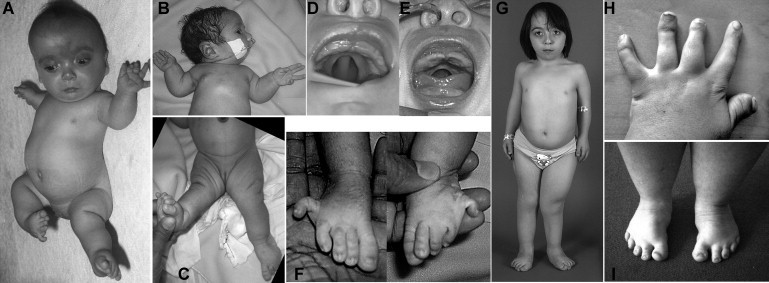
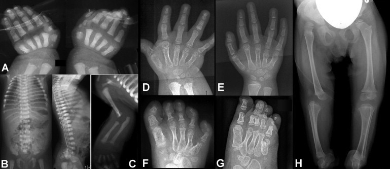
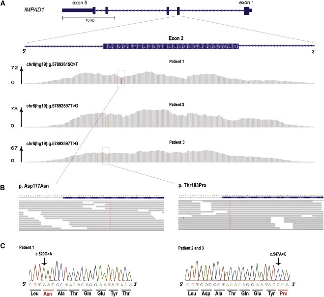
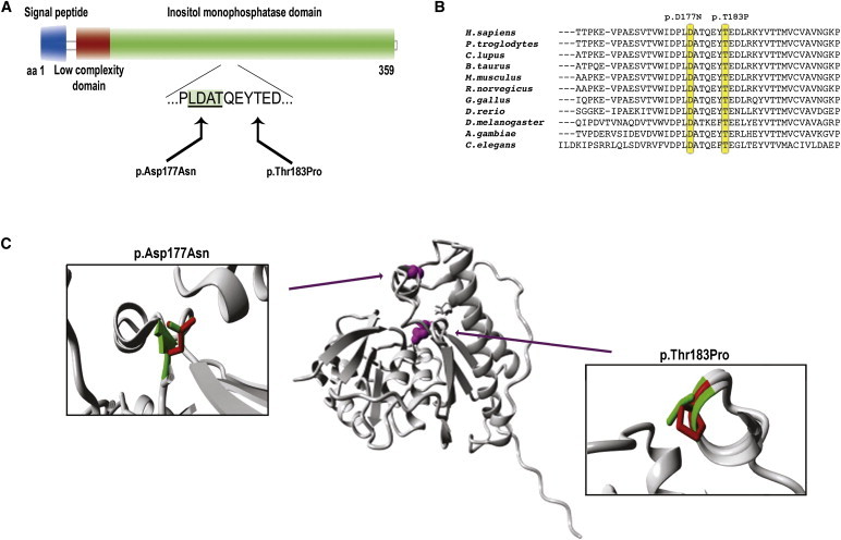
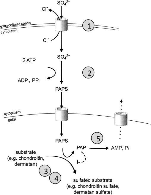
We used whole-exome sequencing to study three individuals with a distinct condition characterized by short stature, chondrodysplasia with brachydactyly, congenital joint dislocations, cleft palate, and facial dysmorphism. Affected individuals carried homozygous missense mutations in IMPAD1, the gene coding for gPAPP, a Golgi-resident nucleotide phosphatase that hydrolyzes phosphoadenosine phosphate (PAP), the byproduct of sulfotransferase reactions, to AMP. The mutations affected residues in or adjacent to the phosphatase active site and are predicted to impair enzyme activity. A fourth unrelated patient was subsequently found to be homozygous for a premature termination codon in IMPAD1. Impad1 inactivation in mice has previously been shown to produce chondrodysplasia with abnormal joint formation and impaired proteoglycan sulfation. The human chondrodysplasia associated with gPAPP deficiency joins a growing number of skeletoarticular conditions associated with defective synthesis of sulfated proteoglycans, highlighting the importance of proteoglycans in the development of skeletal elements and joints.


Figures

Figure 1
Figure 1
Clinical Features of Individuals with IMPAD1 Mutations (A) Patient 2 at age 2 months. A high forehead with a midline hemangioma, a broad nasal bridge, a small mouth, short stature, bilateral anterolateral dislocation of the knees, and lateral clinodactyly of the fifth toes can be seen. (B and C) Patient 4 at age 2 days, with severe retrognathia, limited extension of the elbows, and knee dislocation. (D and E) The median posterior palatal cleft in patients 2 and 4. (F) The proximal position and lateral deviation of the fifth toes in patient 4 (compare with A). (G) Patient 2 at age 9 yrs. Her height was 97 cm and thus far below the 3rd percentile for her age. The hands and feet are short, there are multiple surgical scars, and the right leg is shorter than the left because of hip dislocation. (H and I) Hands and feet of patient 1 at age 23 years. Note the marked brachydactyly associated with shortened metacarpals, as well as the proximal insertion and radial deviation of digit I (so-called “hitch-hiker thumb”). The feet are short, with forefoot adduction.
Figure 2
Figure 2
Radiographic Features of Individuals with IMPAD1 Mutations (A) Hand X-rays of patient 4 at birth show shortening of all metacarpals, the presence of an accessory ossification center at the base of metacarpal 1 (left hand), and splitting of the proximal phalanx of digit I into two parts. (B and C) Findings in patient 4, recorded at birth: there is mild platyspondyly but no coronal clefting, and there is elbow joint dysplasia. (D and E) Hand X-rays of patient 1 (D; age, 2.5 yrs) and patient 2 (E; age, 4 yrs), showing fusion between the hamate and capitate bones, short metacarpals, irregular sizes of the distal metacarpal epiphyses (note the large size of the epiphysis of metacarpal II), and longitudinal splitting of the proximal and distal phalanx of digit 1 (D). The findings are reminiscent of those in Desbuquois dysplasia. (F and G) Foot radiographs of patients 1 and 2, respectively (same ages as the patients in A and B). There are supernumerary tarsal bones; all metatarsal bones are shortened, with metatarsal V being particularly short; note again the difference in size of the distal metatarsal epiphyses in (D). Skeletal age is not accelerated. Clinodactyly of toe V in patient 2 has been corrected by casting. (H) Pelvis and legs of patient 2 at age 3 yrs, showing proximal dislocation of the right femur and persistence of the anterior dislocation of the right knee. Dislocation of the left knee was also present but was not appreciable on this film.
Figure 3
Figure 3
Identification of Homozygous Mutations in IMPAD1 by Exome Sequencing in Patients 1, 2, and 3 (A) Schematic representation of IMPAD1 with detailed overview of exon 2, containing the identified mutations. Note that the coding sequence of IMPAD1 is on the reverse strand of the chromosome 8 sequence, so that the chromosomal sequence corresponds to the noncoding strand. The per-base coverage is provided with an axis to the right indicating the maximum coverage obtained per individual. Coverage represented in gray indicates that the sequence data in the patient showed the wild-type base, whereas colored bases indicate the detection of variants. The relative height of the color is indicative of the presence of variant in the homozygous state. Top: patient 2. Middle: patient 3. Bottom: patient 1. (B) Detailed zoom-in of reads showing the homozygous missense mutations. Grey arrows indicate the mapped sequence reads, with colored bases indicating nucleotides deviating from the wild-type genomic sequence. In the nucleotides of interest, >95% of reads show the mutated sequence. (C) Sanger sequencing validation of the homozygous mutations in the patients 1–3. Note that the orientation is presented from the 5′ to the 3′ end, representing the coding sequence.
Figure 4
Figure 4
Schematic Representation and Protein Modeling of IMPAD1 p.Asp177Asn and p.Thr183Pro Mutations (A) Schematic representation of the predicted effect at the protein level. Domains and functions are provided in different colors, with the binding domain harboring the mutations indicated by an underlined protein sequence in a green shaded box. (B) Evolutionary conservation of the protein sequence throughout evolution. Mutated amino acids are indicated by yellow boxes. (C) Overview of the protein in ribbon presentation. Middle: the protein is colored gray, and the side chains of the mutated residue are colored magenta and shown as small balls. Left and right: Close-up of the p.Thr183Pro and p.Asp177Asn mutations. Side chains of both the wild-type and the mutant residue are shown and are colored green and red, respectively.
Figure 5
Figure 5
Schematic Representation of Sulfate Activation and Sulfation Pathways Extracellular sulfate is taken up from the extracellular space through a sulfate-chloride exchanger (SLC26A2, DTDST) and subsequently activated to its high-energy form, phosophoadenosine phosphosulphate (PAPS). A PAPS translocase that shuttles PAPS from the cytoplasm to the endoplasmic reticulum has been identified in zebrafish. PAPS is the sulfate donor for most sulfotransferase reactions both in the cytoplasm and in the endoplasmic reticulum and Golgi complex, such as those required for sulphation of nascent proteoglycans. Inhibition of sulfotransferase reactions by PAP (dashed line) has been demonstrated in vitro. Efflux of AMP from the endoplasmic reticulum through a transporter or exchanger (dotted line) has been postulated but not demonstrated. Known genetic defects leading to human osteoarticular phenotypes are marked by circled numbers: 1, SLC26A2/DTDST (sulfate transporter); 2, PAPSS (PAPS synthase); 3, CHST3 (chondroitin 6-sulfotransferase); 4, CHST14 (dermatan 4-sulfotransferase); 5, IMPAD1 (Golgi-resident phosphoadenosine phosphate phosphatase, gPAPP).

Similar articles

Cited by

References

    1. Krakow D., Robertson S.P., King L.M., Morgan T., Sebald E.T., Bertolotto C., Wachsmann-Hogiu S., Acuna D., Shapiro S.S., Takafuta T. Mutations in the gene encoding filamin B disrupt vertebral segmentation, joint formation and skeletogenesis. Nat. Genet. 2004;36:405–410. - PubMed
    1. Bicknell L.S., Farrington-Rock C., Shafeghati Y., Rump P., Alanay Y., Alembik Y., Al-Madani N., Firth H., Karimi-Nejad M.H., Kim C.A. A molecular and clinical study of Larsen syndrome caused by mutations in FLNB. J. Med. Genet. 2007;44:89–98. - PMC - PubMed
    1. Rossi A., Superti-Furga A. Mutations in the diastrophic dysplasia sulfate transporter (DTDST) gene (SLC26A2): 22 novel mutations, mutation review, associated skeletal phenotypes, and diagnostic relevance. Hum. Mutat. 2001;17:159–171. - PubMed
    1. Superti-Furga A. Defects in sulfate metabolism and skeletal dysplasias. In: Scriver C.R., Beaudet A.L., Sly W.S., Valle D., Vogelstein B., Childs B., editors. The Metabolic and Molecular Bases of Inherited Disease. Eighth Edition. McGraw-Hill; New York: 2001. pp. 5189–5201.
    1. Hermanns P., Unger S., Rossi A., Perez-Aytes A., Cortina H., Bonafé L., Boccone L., Setzu V., Dutoit M., Sangiorgi L. Congenital joint dislocations caused by carbohydrate sulfotransferase 3 deficiency in recessive Larsen syndrome and humero-spinal dysostosis. Am. J. Hum. Genet. 2008;82:1368–1374. - PMC - PubMed

Publication types

MeSH terms