Case Reports
. 2011 May 13;88(5):635-42.
doi: 10.1016/j.ajhg.2011.04.006. Epub 2011 May 5.

Exome sequencing identifies mitochondrial alanyl-tRNA synthetase mutations in infantile mitochondrial cardiomyopathy

Affiliations
Case Reports

Exome sequencing identifies mitochondrial alanyl-tRNA synthetase mutations in infantile mitochondrial cardiomyopathy

Alexandra Götz et al. Am J Hum Genet. .

Abstract

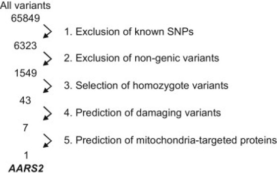
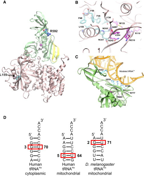
Infantile cardiomyopathies are devastating fatal disorders of the neonatal period or the first year of life. Mitochondrial dysfunction is a common cause of this group of diseases, but the underlying gene defects have been characterized in only a minority of cases, because tissue specificity of the manifestation hampers functional cloning and the heterogeneity of causative factors hinders collection of informative family materials. We sequenced the exome of a patient who died at the age of 10 months of hypertrophic mitochondrial cardiomyopathy with combined cardiac respiratory chain complex I and IV deficiency. Rigorous data analysis allowed us to identify a homozygous missense mutation in AARS2, which we showed to encode the mitochondrial alanyl-tRNA synthetase (mtAlaRS). Two siblings from another family, both of whom died perinatally of hypertrophic cardiomyopathy, had the same mutation, compound heterozygous with another missense mutation. Protein structure modeling of mtAlaRS suggested that one of the mutations affected a unique tRNA recognition site in the editing domain, leading to incorrect tRNA aminoacylation, whereas the second mutation severely disturbed the catalytic function, preventing tRNA aminoacylation. We show here that mutations in AARS2 cause perinatal or infantile cardiomyopathy with near-total combined mitochondrial respiratory chain deficiency in the heart. Our results indicate that exome sequencing is a powerful tool for identifying mutations in single patients and allows recognition of the genetic background in single-gene disorders of variable clinical manifestation and tissue-specific disease. Furthermore, we show that mitochondrial disorders extend to prenatal life and are an important cause of early infantile cardiac failure.


Figures

Figure 1
Figure 1
The Mitochondrial Cardiomyopathy Patients and Their Families (A) Pedigrees of families 1 and 2. (B) Cytochrome c oxidase (COX) activity (shown in brown) of heart and skeletal muscle of patients 1 (II-1, family 1) and 2 (II-6, family 2). Simultaneous histochemical analysis for COX and succinate dehydrogenase (SDH, shown in blue) activities (Cox/Sdh) on frozen cryostat sections revealed mitochondrial COX deficiency with mitochondrial proliferation. (C) Blue native electrophoresis analyses of mitochondrial respiratory chain complexes in the heart, brain, and liver of patient 1 (P1), compared to control samples (c). Ten micrograms of sample protein was loaded onto a gel. For protein detection, monoclonal antibodies (Mitosciences) against the 39 kDa subunit of Complex I (CI), the subunits core 2 or Rieske of Complex III (CIII), the cox1p and cox4 subunits of Complex IV (CIV), and the 70 kDa-Ip subunit of Complex II (CII) were used.
Figure 2
Figure 2
Schematic Representation of the Exome Data Analysis and Data Filtering (1) The known dbSNP130 variants were excluded assuming the pathogenic variant to be too rare to (yet) exist in the database. (2) Nongenic variants were excluded. (3) Homozygous changes were selected: autosomal infantile-onset mitochondrial disorders are generally recessively inherited and homozygosity of the pathogenic variant was considered possible as the patient originated from Finland, a genetic isolate. (4) The consequence of the remaining variants was assessed by SIFT Genome tool (5) Genes encoding proteins with a mitochondrial function were predicted by MitoProt and TargetP.
Figure 3
Figure 3
AARS2 and Mutations (A) AARS2 mutation sequences in patients 1 (II-1, family 1) and 2 (II-6, family 2). (B) Cross-species protein conservation of mtAlaRS, flanking the altered amino acids p.Leu155Arg and p.Arg592Trp in mammals. The corresponding gene has not been fully characterized in other vertebrate species. (C) Schematic representation of AARS2. Boxes represent exons 1–22. Two main functional domains, the aminoacylation (pink) and editing (green) domains, are indicated. M denotes the mitochondrial targeting signal. The p.Leu155Arg mutation is located in the aminoacylation domain, p.Arg592Trp in the editing domain. (D) The AARS2-GFP fusion protein (green) colocalizes with Mitotracker Red (Invitrogen), indicating mitochondrial localization for mtAlaRS in HEK293T cells (overlay in yellow). The cells were examined and imaged with an Olympus IX8 fluorescence microscope.
Figure 4
Figure 4
Modeled Human mtAlaRS (A) The location of Leu155 within the aminoacylation domain (pink) and of Arg592 within the editing domain (green). The strand-loop-strand motif (aa 764–783) in the editing domain is shown in yellow. (B) Hydrophobic architectural residues surrounding the catalytic aminoacylation site. Carbon atoms of the catalytic residues involved in amino acid binding and aminoacyl adenylate formation are shown in magenta. (C) Conserved positively charged residues, predicted to be involved in the recognition and binding of tRNA (yellow) in the editing domain of human mtAlaRS. (D) Comparative analysis of the predicted secondary structures of human cytoplasmic and mitochondrial tRNAAla revealed that the identity base pair of the latter is likely to be G5:U64, implying a structurally unique tRNA recognition site in the human mtAlaRS.
Figure 5
Figure 5
Metabolomic Analysis: Postmortem Heart and Skeletal Muscle of Patient Shows Increased Alanine Levels Patient 1 (II-1, family 1) heart, muscle, and liver samples were compared to those of an age-matched patient with nonmitochondrial dilated CMP (heart and skeletal muscle) and those of a 10-year-old patient with mitochondrial encephalopathy (heart and liver).

References

    1. Thorburn D.R. Mitochondrial disorders: prevalence, myths and advances. J. Inherit. Metab. Dis. 2004;27:349–362. - PubMed
    1. Holmgren D., Wåhlander H., Eriksson B.O., Oldfors A., Holme E., Tulinius M. Cardiomyopathy in children with mitochondrial disease; clinical course and cardiological findings. Eur. Heart J. 2003;24:280–288. - PubMed
    1. Scaglia F., Towbin J.A., Craigen W.J., Belmont J.W., Smith E.O., Neish S.R., Ware S.M., Hunter J.V., Fernbach S.D., Vladutiu G.D. Clinical spectrum, morbidity, and mortality in 113 pediatric patients with mitochondrial disease. Pediatrics. 2004;114:925–931. - PubMed
    1. Yaplito-Lee J., Weintraub R., Jamsen K., Chow C.W., Thorburn D.R., Boneh A. Cardiac manifestations in oxidative phosphorylation disorders of childhood. J. Pediatr. 2007;150:407–411. - PubMed
    1. Tanaka M., Ino H., Ohno K., Hattori K., Sato W., Ozawa T., Tanaka T., Itoyama S. Mitochondrial mutation in fatal infantile cardiomyopathy. Lancet. 1990;336:1452. - PubMed

Publication types