. 2011 Aug 31;665(1-3):19-28.
doi: 10.1016/j.ejphar.2011.04.039. Epub 2011 Apr 28.

Chronic type II diabetes mellitus leads to changes in neuropeptide Y receptor expression and distribution in human myocardial tissue

Affiliations

Chronic type II diabetes mellitus leads to changes in neuropeptide Y receptor expression and distribution in human myocardial tissue

Robina Matyal et al. Eur J Pharmacol. .

Abstract

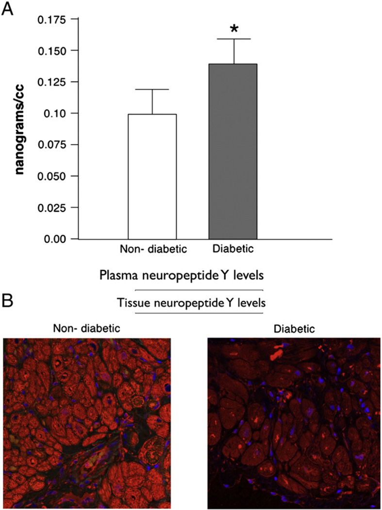
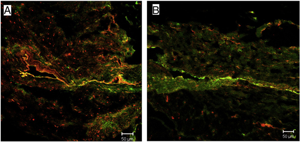
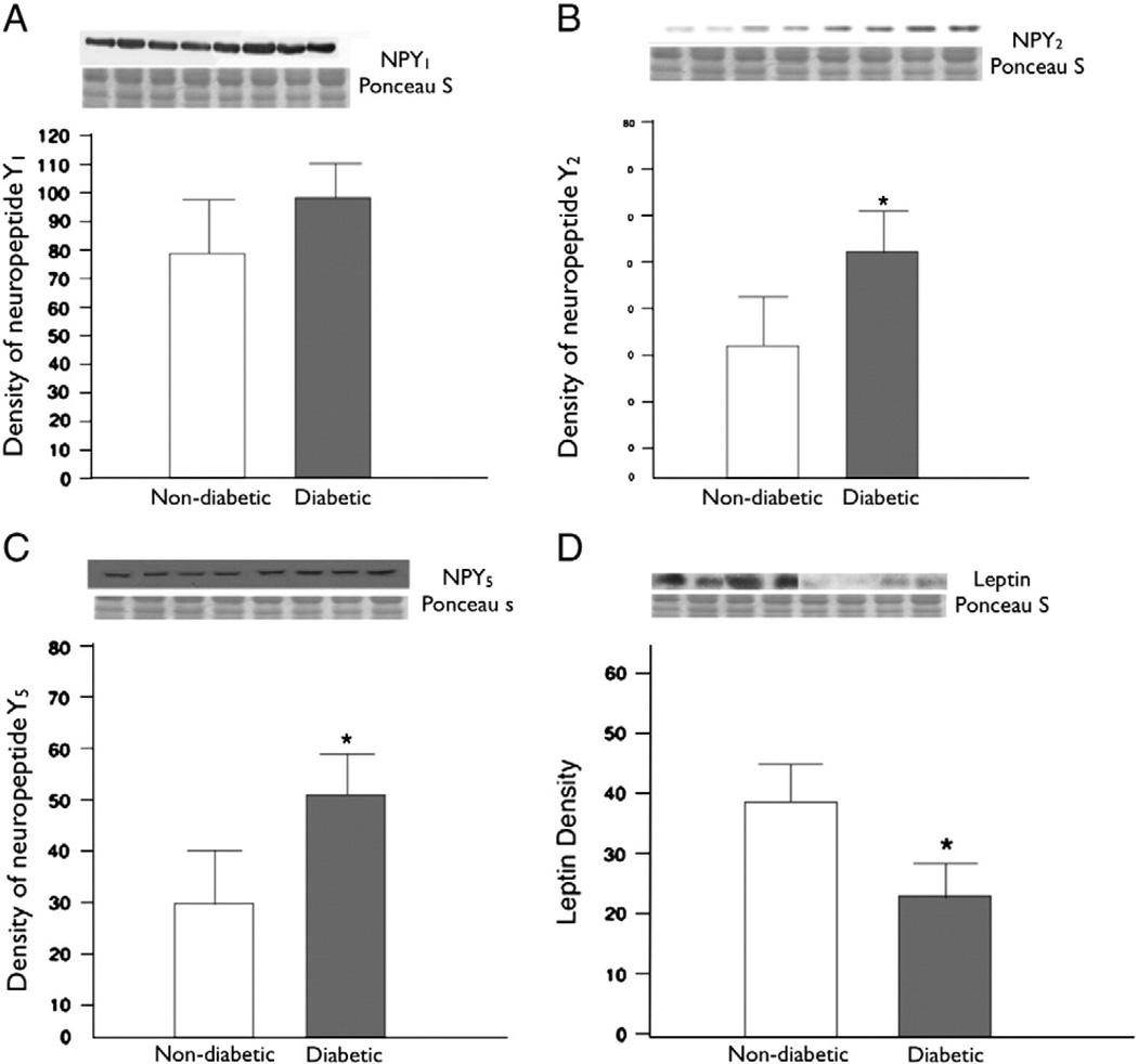
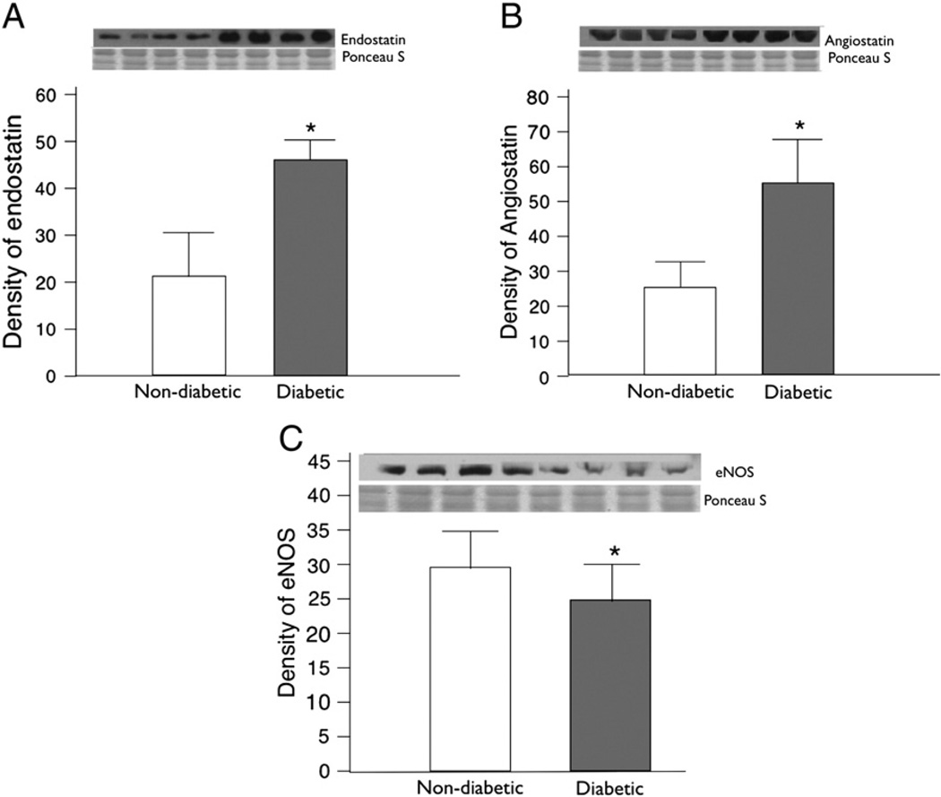
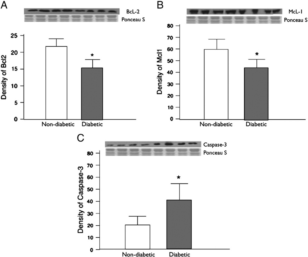
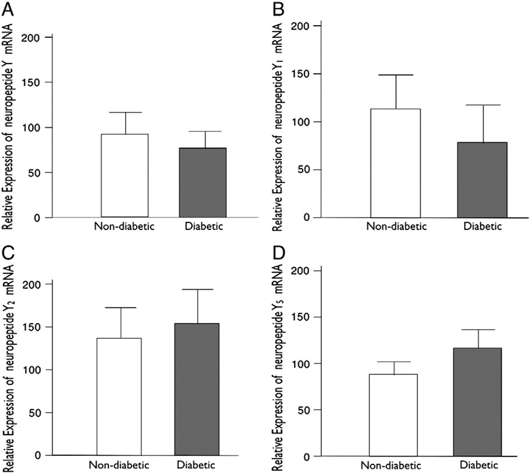
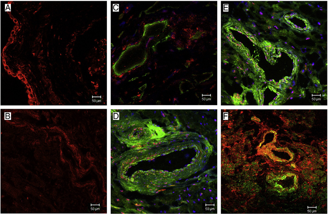
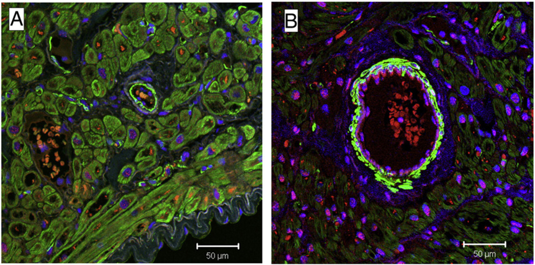
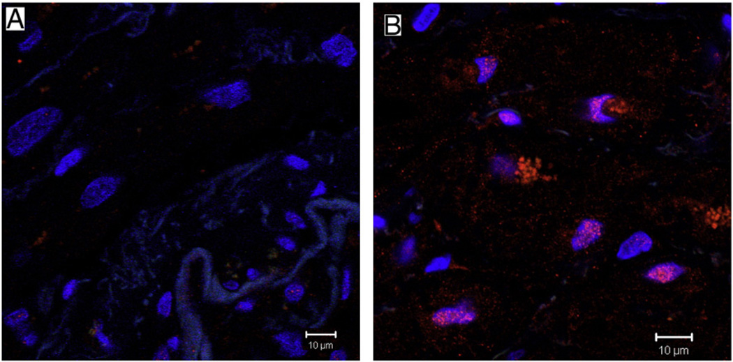
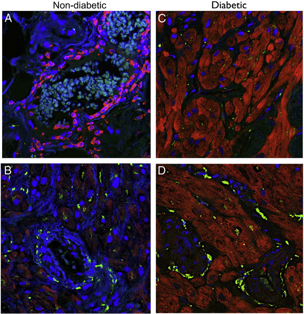
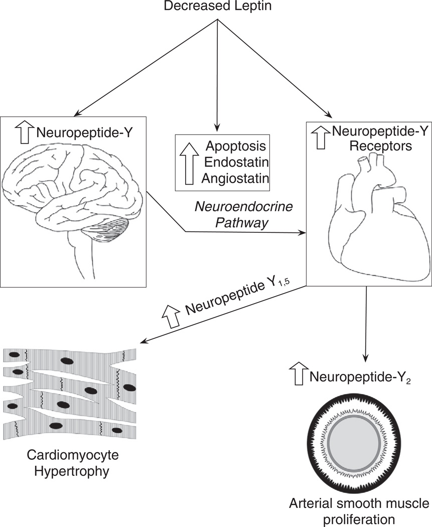
Neuropeptide Y is one of the most abundant neurotransmitters in the myocardium, and is known to influence cardiovascular remodeling. We hypothesized that diabetic neuropathy could possibly be associated with altered neuropeptide Y and its receptor expression levels in myocardium and plasma. Plasma neuropeptide Y levels in diabetic (n=24, HgbA1c 7.9 ± 1.1%) and non-diabetic (n=27, HgbA1c 5.8 ± 0.5%) patients undergoing cardiac surgery utilizing cardiopulmonary bypass were analyzed. Right atrial tissue of these patients was used to determine the expression of neuropeptide Y, the receptors 1-5, and leptin by immunoblotting, real-time PCR and immunofluorescence. Apoptosis signaling and endostatin and angiostatin were measured to determine the effects of leptin. Plasma neuropeptide Y levels were significantly increased in patients with Type II diabetes mellitus as compared to non-diabetic patients (P=0.026). Atrial tissue neuropeptide Y mRNA levels were lower in diabetic patients (P=0.036). There was a significant up-regulation of myocardial Y(2) and Y(5) receptors (P=0.009, P=0.01 respectively) in the diabetic patients. Leptin, involved with apoptosis and angiogenesis, was down regulated in diabetic patients (P=0.05). The levels of caspase-3, endostatin and angiostatin were significantly elevated in diabetic patients (P=0.003, P=0.008, P=0.01 respectively). Y(1) receptors were more likely to be localized within the nuclei of cardiomyocytes and vascular smooth muscle cells. Neuropeptide expression is altered differentially in the serum and myocardium by diabetes. Altered regulation of this system in diabetics may be in part responsible for the decreased angiogenesis, increased apoptosis, and increased vascular smooth muscle proliferation leading to coronary artery disease and heart failure in this patient population.


Figures

Fig. 1
Fig. 1
(A) Plasma levels of neuropeptide Y in non-diabetic with HbA1c levels <5.8% versus diabetic with HbA1c levels >6.8% patients as measured by ELISA immunoassay. The plasma levels were significantly elevated in diabetic patients as compared to non-diabetic patients (P=0.026). (B) Immunohistochemistry staining with neuropeptide Y showing decreased levels of neuropeptide Y in diabetic tissue as compared to non-diabetic atrial tissue.
Fig. 2
Fig. 2
Representative confocal microscopy. neuropeptide Y (red) co-stained with the general nerve marker protein gene product 9.5 (PGP 9.5, green), showing neuropeptide Y staining in perivascular nerves in non-diabetic and diabetic patient atrial tissue. There is decreased neuropeptide Y co-staining with PGP 9.5 in diabetic atrial tissue (B) as compared to non-diabetic tissue (A).
Fig. 3
Fig. 3
Protein expression was assessed in the atrial tissue of non-diabetic and diabetic patients. (A) neuropeptide Y1 levels were elevated but not significantly, in diabetic patients than in non-diabetic patients (P=0.2). (B) neuropeptide Y2 were significantly elevated in diabetic patients than in non-diabetic patients (P=0.008). (C) neuropeptide Y5 were significantly elevated in diabetic patients than in non-diabetics (P=0.01). (D) Leptin protein was significantly reduced in diabetic patients as compared to non-diabetics, (P=0.05).
Fig. 4
Fig. 4
(A) Endostatin levels were assessed in the atrial tissue, the endostatin levels were significantly elevated in non-diabetic as compared to diabetic (P=0.008). (B) The levels of angiostatin were significantly elevated in diabetic as compared to non-diabetic patients (P=0.01). (C) The expression of eNOS was decreased in diabetic as compared to non-diabetic (P=0.01).
Fig. 5
Fig. 5
(A) The levels of pro-survival factor BCl-2 were decreased in diabetic as compared to non-diabetic (P=0.003) (B) The levels of another pro-survival factor MCl-1 were significantly decreased in diabetic as compared to non-diabetic (P=0.003) (C) There were significantly elevated levels of caspase-3 in diabetic as compared to non-diabetic (P=0.003).
Fig. 6
Fig. 6
(A) Myocardial levels of NPY mRNA in non-diabetic and diabetic patients as measured by semi-quantitative real-time PCR (P=0.36). (B) Myocardial levels of neuropeptide Y1 mRNA in non-diabetic (n=12) versus diabetic (n=12) patients as measured by real-time PCR (P=0.7). (C) Relative expression of neuropeptide Y2 in diabetic patients as compared to non-diabetic patients (P=0.67). (D) Relative expression of neuropeptide Y5 in diabetic patients as compared to non-diabetics (P=0.5).
Fig. 7
Fig. 7
Representative immunofluorescence staining for Y1 (red) in sections co-stained with the vascular smooth muscle marker -smooth muscle actin (green) in atrial tissue from Non-diabetic and Diabetic patients. (A and B) There is staining of Y1 receptor in the endothelium of non-diabetic tissue, while the staining is decreased in the endothelium of diabetic tissue. (D and F) There are increased staining of Y1 receptor around the arteries in the atrial tissue of diabetic patients as compared to non-diabetic patients (C and E).
Fig. 8
Fig. 8
Immunofluorescence images. Representative slides of atrial tissue from non-diabetic and diabetic patients. Sections were co-stained with 4′,6′-diamidino-2-phenylindole (DAPI, blue) to demarcate the nucleus, green for alpha smooth muscle actin and red for Y1 receptor. There is increased Y1 receptor expression around the arteries and in the cardiomyocyte nuclei in diabetic patient. Magnification for both images is 20×.
Fig. 9
Fig. 9
Sections were co-stained with 4′,6′-diamidino-2-phenylindole (DAPI, blue) to demarcate the nucleus. Magnification for all images is 63×. The number of nuclei staining with Y1 nuclear staining+cells/mm2) is statistically significant in diabetic patients (B) as compared to non-diabetics (A) (P=0.04).
Fig. 10
Fig. 10
Immunofluorescence images. Representative slides of atrial tissue from non-diabetic and diabetic patients. Sections were co-stained with 4′,6′-diamidino-2-phenylindole (DAPI, blue) to demarcate the nucleus and Y5 receptors. Magnification for all images is 40×. There is increased staining for Y5 receptors in diabetic tissue cardiomyocytes (C and D) as compared to non-diabetic tissue where the localization of Y5 receptors is around blood vessel. (A and B).
Fig. 11
Fig. 11
Schematic diagram of leptin interaction and neuropeptide Y. In diabetes mellitus there is either decreased leptin or leptin resistance that affects neuropeptide Y levels in the hypothalamus and peripheral autonomic nervous system. This altered regulation of neuropeptide Y leads to changes in neuropeptide Y receptor concentration. Decreased leptin itself causes increased apoptosis and increased anti-angiogenic factors. The neuropeptide Y receptorsmay be involved in cardiomyocytes hypertrophy and arterial smoothmuscle proliferation.

References

    1. Abe K, Tilan JU, Zukowska Z. NPY and NPY receptors in vascular remodeling. Curr. Top. Med. Chem. 2007;7:1704–1709. - PubMed
    1. Abe K, Kuo L, Zukowska Z. Neuropeptide Y is a mediator of chronic vascular and metabolic maladaptations to stress and hypernutrition. Exp. Biol. Med. (Maywood) 2010;235:1179–1184. - PubMed
    1. Chottova Dvorakova M, Wiegand S, Pesta M, Slavikova J, Grau V, Reischig J, Kuncova J, Kummer W. Expression of neuropeptide Y and its receptors Y1 and Y2 in the rat heart and its supplying autonomic and spinal sensory ganglia in experimentally induced diabetes. Neuroscience. 2008;151:1016–1028. - PubMed
    1. Fang ZY, Prins JB, Marwick TH. Diabetic cardiomyopathy: evidence, mechanisms, and therapeutic implications. Endocr. Rev. 2004;25:543–567. - PubMed
    1. Gu J, Polak JM, Allen JM, Huang WM, Sheppard MN, Tatemoto K, Bloom SR. High concentrations of a novel peptide, neuropeptide Y, in the innervation of mouse and rat heart. J. Histochem. Cytochem. 1984;32:467–472. - PubMed

Publication types

Substances