. 2011 Jun 24;409(5):853-66.
doi: 10.1016/j.jmb.2011.04.044. Epub 2011 Apr 27.

Design and characterization of epitope-scaffold immunogens that present the motavizumab epitope from respiratory syncytial virus

Affiliations

Design and characterization of epitope-scaffold immunogens that present the motavizumab epitope from respiratory syncytial virus

Jason S McLellan et al. J Mol Biol. .

Abstract

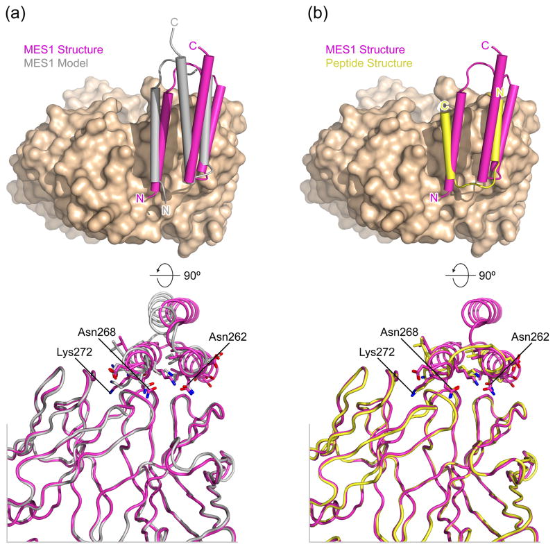
Respiratory syncytial virus (RSV) is a major cause of respiratory tract infections in infants, but an effective vaccine has not yet been developed. An ideal vaccine would elicit protective antibodies while avoiding virus-specific T-cell responses, which have been implicated in vaccine-enhanced disease with previous RSV vaccines. We propose that heterologous proteins designed to present RSV-neutralizing antibody epitopes and to elicit cognate antibodies have the potential to fulfill these vaccine requirements, as they can be fashioned to be free of viral T-cell epitopes. Here we present the design and characterization of three epitope-scaffolds that present the epitope of motavizumab, a potent neutralizing antibody that binds to a helix-loop-helix motif in the RSV fusion glycoprotein. Two of the epitope-scaffolds could be purified, and one epitope-scaffold based on a Staphylococcus aureus protein A domain bound motavizumab with kinetic and thermodynamic properties consistent with the free epitope-scaffold being stabilized in a conformation that closely resembled the motavizumab-bound state. This epitope-scaffold was well folded as assessed by circular dichroism and isothermal titration calorimetry, and its crystal structure (determined in complex with motavizumab to 1.9 Å resolution) was similar to the computationally designed model, with all hydrogen-bond interactions critical for binding to motavizumab preserved. Immunization of mice with this epitope-scaffold failed to elicit neutralizing antibodies but did elicit sera with F binding activity. The elicitation of F binding antibodies suggests that some of the design criteria for eliciting protective antibodies without virus-specific T-cell responses are being met, but additional optimization of these novel immunogens is required.


Figures

Fig. 1
Fig. 1. Motavizumab epitope-scaffolds
Three proteins were computationally designed to accept and maintain the motavizumab epitope in the conformation observed in the complex between motavizumab and its epitope peptide. (a) Ribbon diagram of a pre-fusion RSV F model, and the structure of the motavizumab epitope peptide bound to motavizumab Fab. The motavizumab heavy chain is green, and the light chain is blue. The epitope peptide is grey, with the 13 residues that were transplanted into the acceptor-scaffolds colored red. (b) Models of the three motavizumab epitope-scaffolds, in an orientation similar to that of the peptide in (a). The transplanted residues are colored red, and the PDB ID and chain ID of the original structures are listed.
Fig. 2
Fig. 2. Kinetics and thermodynamics of epitope-scaffolds binding to motavizumab
MES1 has a faster on-rate than the peptide, and MES1 binding is entropically driven, suggesting that the motavizumab epitope present on MES1 is fixed in the bound-conformation. (a–c) SPR sensorgrams of MES1, MES2 and F peptide binding to immobilized motavizumab Fab. Red lines represent best fit of kinetic data to 1:1 binding models. (d–f) ITC data for MES1, MES2 and F peptide binding to motavizumab IgG. Experiments were performed at 25ºC in PBS, and red lines represent best fit of data to a single binding-site model.
Fig. 3
Fig. 3. Circular dichroism data for MES1 and MES2
Both epitope-scaffolds have spectra indicative of alpha-helical proteins, but only MES1 displays cooperative-unfolding. (a) MES1 CD spectrum. (b) MES1 melting profile obtained by measuring mean residue ellipticity at 210 nm while increasing the temperature from 5ºC to 99ºC. Red line represents best fit of the data to a two-state model. (c) MES2 CD spectrum. (d) MES2 melting profile obtained by measuring mean residue ellipticity at 210 nm while increasing the temperature from 5ºC to 99ºC. The non-sigmoidal plot indicates a lack of cooperative unfolding, and the data could not be fit to a two-state model.
Fig. 4
Fig. 4. Crystal structure of MES1 bound to motavizumab
The conformation of motavizumab-bound MES1 is similar to the motavizumab-bound peptide and the computationally-designed model. (a) Structure of MES1 in complex with motavizumab Fab. A semi-transparent molecular surface is overlaid on a ribbon representation of the complex. The motavizumab heavy chain is green, the light chain is blue, and MES1 is magenta. (b) Cα superposition of the 13 shared residues between MES1 (magenta) and the F peptide (yellow) in ribbon representation. The rmsd is 0.30 A for the 13 Cα residues. (c) Cα superposition of MES1 from the crystal structure (magenta) and the model (grey) in ribbon representation. The rmsd is 1.16 Å for 53 Cα residues. (d) Plot of the Cα distances of the superposition shown in (c). The 20 helical residues which contain the 13 transplanted residues are colored red.
Fig. 5
Fig. 5. MES1 binding to motavizumab Fab
MES1 binding to motavizumab is shifted with respect to the model and the epitope peptide, though side-chain hydrogen-bond interactions are preserved. (a) MES1 in complex with motavizumab based on coordinates from the crystal structure (magenta) and the model (grey). Superposition of the Fab molecules was used to orient the complexes. The top image shows the molecular surface of the Fab, and MES1 alpha-helices are depicted as cylinders. The bottom image shows both MES1 and the Fab as ribbons, with the side-chains of the 13 transplanted residues shown as sticks. Oxygen atoms are colored red and nitrogen atoms are blue. (b) Same as (a), except the peptide/motavizumab crystal structure (yellow) replaces the MES1/motavizumab model.
Fig. 6
Fig. 6. Epitope-scaffold immunogenicity
MES1 elicits antibodies when fused to a PADRE sequence, and sera with F protein-binding activity are elicited by combinations of MES1, a modified derivative of MES1, or MES2. (a) ELISA analysis of sera from MES1-immunized mice. MES1 or a mutant that ablates motavizumab binding (MES1 K272E), were coated on the plate. (b,c) Sera from mice immunized with epitope-scaffolds were tested for binding to MES1 (b) and recombinant RSV F protein (c). (d) Sera from mice immunized four times with MES1 were tested for binding to proteins that had been pre-incubated with motavizumab IgG. For all panels, plotted data represent the mean of 5 mice ± SD. As a reference, a result of 100 mOD/min in the kinetic ELISA is equivalent to the F binding activity of ~0.5 μg/ml of palivizumab (Synagis®, Medimmune, Gaithersburg, MD) and a result of 20 mOD/min ~0.01 μg/ml.
Fig. 7
Fig. 7. MES1 derivative for focusing the immune response
Ribbon diagram of MES1 model (a), with residues replaced in Surf1 (b) shown as sticks with semi-transparent surfaces. (c) SPR sensorgram of Surf1 binding to immobilized motavizumab Fab. Five 2-fold dilutions of Surf1 starting at 500 nM were measured. Red lines represent best fit of kinetic data to a 1:1 binding model.

References

    1. Hall CB, Weinberg GA, Iwane MK, Blumkin AK, Edwards KM, Staat MA, Auinger P, Griffin MR, Poehling KA, Erdman D, Grijalva CG, Zhu Y, Szilagyi P. The Burden of Respiratory Syncytial Virus Infection in Young Children. N Engl J Med. 2009;360:588–598. - PMC - PubMed
    1. Nair H, Nokes DJ, Gessner BD, Dherani M, Madhi SA, Singleton RJ, O'Brien KL, Roca A, Wright PF, Bruce N, Chandran A, Theodoratou E, Sutanto A, Sedyaningsih ER, Ngama M, Munywoki PK, Kartasasmita C, Simões EAF, Rudan I, Weber MW, Campbell H. Global burden of acute lower respiratory infections due to respiratory syncytial virus in young children: a systematic review and meta-analysis. The Lancet. 2010;375:1545–1555. - PMC - PubMed
    1. Kim HW, Canchola JG, Brandt CD, Pyles G, Chanock RM, Jensen K, Parrott RH. Respiratory Syncytial Virus Disease in Infants Despite Prior Administration of Antigenic Inactivated Vaccine. Am J Epidemiol. 1969;89:422–434. - PubMed
    1. Graham BS. Biological challenges and technological opportunities for respiratory syncytial virus vaccine development. Immunol Rev. 2011;239:149–66. - PMC - PubMed
    1. Graham B, Henderson G, Tang Y, Lu X, Neuzil K, Colley D. Priming immunization determines T helper cytokine mRNA expression patterns in lungs of mice challenged with respiratory syncytial virus. J Immunol. 1993;151:2032–2040. - PubMed

Publication types

Associated data