. 2011 Sep;1812(9):1182-9.
doi: 10.1016/j.bbadis.2011.04.006. Epub 2011 Apr 27.

Assessment of newly synthesized mitochondrial DNA using BrdU labeling in primary neurons from Alzheimer's disease mice: Implications for impaired mitochondrial biogenesis and synaptic damage

Affiliations

Assessment of newly synthesized mitochondrial DNA using BrdU labeling in primary neurons from Alzheimer's disease mice: Implications for impaired mitochondrial biogenesis and synaptic damage

Marcus J Calkins et al. Biochim Biophys Acta. 2011 Sep.

Abstract

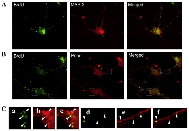
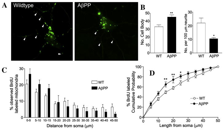
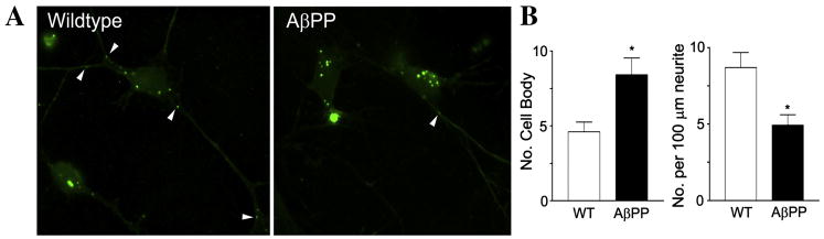
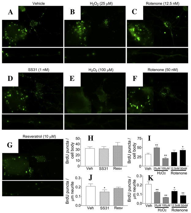
The purpose of our study was to assess mitochondrial biogenesis and distribution in murine primary neurons. Using 5-bromo-2-deoxyuridine (BrdU) incorporation and primary neurons, we studied the mitochondrial biogenesis and mitochondrial distribution in hippocampal neurons from amyloid beta precursor protein (AβPP) transgenic mice and wild-type (WT) neurons treated with oxidative stressors, rotenone and H(2)O(2). We found that after 20h of labeling, BrdU incorporation was specific to porin-positive mitochondria. The proportion of mitochondrial area labeled with BrdU was 40.3±6.3% at 20h. The number of mitochondria with newly synthesized DNA was higher in AβPP neuronal cell bodies than in the cell bodies of WT neurons (AβPP, 45.23±2.67 BrdU-positive/cell body; WT, 32.92±2.49 BrdU-positive/cell body; p=0.005). In neurites, the number of BrdU-positive mitochondria decreased in AβPP cultures compared to WT neurons (AβPP, 0.105±0.008 BrdU-positive/μm neurite; WT, 0.220±0.036 BrdU-positive/μm neurite; p=0.010). Further, BrdU in the cell body increased when neurons were treated with low doses of H(2)O(2) (49.6±2.7 BrdU-positive/cell body, p=0.0002 compared to untreated cells), while the neurites showed decreased BrdU staining (0.122±0.010 BrdU-positive/μm neurite, p=0.005 compared to the untreated). BrdU labeling was increased in the cell body under rotenone treatment. Additionally, under rotenone treatment, the content of BrdU labeling decreased in neurites. These findings suggest that Aβ and mitochondrial toxins enhance mitochondrial fragmentation in the cell body, and may cause impaired axonal transport of mitochondria leading to synaptic degeneration.


Figures

Figure 1
Figure 1. BrdU staining is localized to mitochondria in primary neuronal cultures
Primary hippocampal neurons were incubated with BrdU for 20 hrs and then stained to visualize BrdU incorporation. Cells were then co-labeled with MAP-2 (neuronal marker) or porin (mitochondrial marker). Primary neurons as identified by MAP-2 staining (A - upper panels) showed robust BrdU labeling as puncta within the cell body and neurites. These BrdU puncta co-localized with porin (B - middle panels), indicating that BrdU was incorporated into the mtDNA. Enlargements (C – lower panels) show co-localization of BrdU is specific to porin-stained organelles (arrowheads). Enlargements represent a, BrdU; b, porin; c, merged; d, BrdU; e, Porin; f, Merged.
Figure 2
Figure 2. AβPP primary hippocampal neurons show increased BrdU staining in the cell body and decreased BrdU staining in neurites
Primary neurons grown from AβPP and wild-type mice were incubated with BrdU for 20 hr at 10 DIV and then stained for analysis. Representative images of AβPP and WT neurons are shown (A). Quantification of BrdU-labeled mitochondria in the cell body and neruites is shown (B). Quantification of BrdU puncta distribution in relation to the cell soma was performed and analyzed by two methods. The proportion of BrdU-stained puncta in 5-μm bins is shown (C), while the proportion of BrdU puncta within a certain length of neuritic distance is also shown (D). A higher proportion of BrdU-labeled mitochondria were in close proximity to the cell body in the AβPP cultures compared to wild-type cultures.
Figure 3
Figure 3. Primary neurons from AβPP mice showed altered BrdU distribution even after short-term BrdU incubation
Representative images of AβPP primary hippocampal neurons that were incubated with BrdU for 4 hr are shown (A). Quantification of cell body and BrdU puncta in neurites are shown (B).
Figure 4
Figure 4. Both oxidative toxins and neuroprotective molecules alter the distribution of BrdU-stained mitochondria
Wild-type cells treated with neuroprotective molecules SS31 (D) and resveratrol (G) for 48 hr, or with H2O2 (B & E) and rotenone (F) for 24 hr were assayed for BrdU incorporation into mitochondria. Representative images for all treatment groups, enlargements of the cell body and selected neurites are shown. SS31 treatment resulted in no change to BrdU staining in the cell body (D & H), but it did result in reduced BrdU staining in the neurites (G,H &J). Images show resveratrol treatment did not alter BrdU staining in the cell body or the neurites. A mildly toxic dose of H2O2 (25 μM) caused an accumulation of BrdU puncta in the cell body and a reduction in the neuritic processes (B,I &K). A higher dose (100 μM) resulted in the loss of BrdU staining in both the cell body and neurites. Two doses of rotenone that did not cause overt toxicity in the culture resulted in an accumulation of BrdU staining in the cell body and a dose-dependent decrease in BrdU staining, in neurites (C,F,I & K). Quantification of images from multiple cultures is shown (H,I,J &K). * p < 0.05 compared to the vehicle-treated group. ** p < 0.005 compared to the vehicle-treated group.

References

    1. Beal MF. Mitochondria take center stage in aging and neurodegeneration. Ann Neurol. 2005;58:495–505. - PubMed
    1. Keller JN, Pang Z, Geddes JW, Begley JG, Germeyer A, Waeg G, Mattson MP. Impairment of glucose and glutamate transport and induction of mitochondrial oxidative stress and dysfunction in synaptosomes by amyloid beta-peptide: role of the lipid peroxidation product 4-hydroxynonenal. J Neurochem. 1997;69:273–84. - PubMed
    1. Reddy PH. Mitochondrial medicine for aging and neurodegenerative diseases. Neuromolecular Med. 2008;10:291–315. - PMC - PubMed
    1. Swerdlow RH, Khan SM. A “mitochondrial cascade hypothesis” for sporadic Alzheimer’s disease. Med Hypotheses. 2004;63:8–20. - PubMed
    1. Reddy PH, Beal MF. Amyloid beta, mitochondrial dysfunction and synaptic damage: implications for cognitive decline in aging and Alzheimer’s disease. Trends Mol Med. 2008;14:45–53. - PMC - PubMed

Publication types