. 2011 Jul 1;22(13):2258-69.
doi: 10.1091/mbc.E10-12-0934. Epub 2011 May 5.

The two actin-interacting protein 1 genes have overlapping and essential function for embryonic development in Caenorhabditis elegans

Affiliations

The two actin-interacting protein 1 genes have overlapping and essential function for embryonic development in Caenorhabditis elegans

Shoichiro Ono et al. Mol Biol Cell. .

Abstract

Disassembly of actin filaments by actin-depolymerizing factor (ADF)/cofilin and actin-interacting protein 1 (AIP1) is a conserved mechanism to promote reorganization of the actin cytoskeleton. We previously reported that unc-78, an AIP1 gene in the nematode Caenorhabditis elegans, is required for organized assembly of sarcomeric actin filaments in the body wall muscle. unc-78 functions in larval and adult muscle, and an unc-78-null mutant is homozygous viable and shows only weak phenotypes in embryos. Here we report that a second AIP1 gene, aipl-1 (AIP1-like gene-1), has overlapping function with unc-78, and that depletion of the two AIP1 isoforms causes embryonic lethality. A single aipl-1-null mutation did not cause a detectable phenotype. However, depletion of both unc-78 and aipl-1 arrested development at late embryonic stages due to severe disorganization of sarcomeric actin filaments in body wall muscle. In vitro, both AIPL-1 and UNC-78 preferentially cooperated with UNC-60B, a muscle-specific ADF/cofilin isoform, in actin filament disassembly but not with UNC-60A, a nonmuscle ADF/cofilin. AIPL-1 is expressed in embryonic muscle, and forced expression of AIPL-1 in adult muscle compensated for the function of UNC-78. Thus our results suggest that enhancement of actin filament disassembly by ADF/cofilin and AIP1 proteins is critical for embryogenesis.


Figures

FIGURE 1:
FIGURE 1:
AIPL-1 is a second AIP1 isoform in C. elegans. AIPL-1 and UNC-78 from C. elegans are 66% identical in their amino acid sequences. Sequence alignment is shown in Supplemental Figure S1. A phylogenetic tree of AIP1 sequences from various species was generated by the neighbor-joining method using CLC Sequence Viewer (CLC Bio, Cambridge, MA).
FIGURE 2:
FIGURE 2:
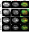
Expression patterns of aipl-1 and unc-78. The promoter region of aipl-1 or unc-78 was fused with a GFP reporter, and the patterns of GFP expression were examined. (A–L) Promoter activities of aipl-1 (A–C, G–I) and unc-78 (D–F, J–L) in embryos. Embryos were fixed and stained with anti-GFP (A, D, G, J) and anti–MYO-3 (a marker for the body wall muscle) (B, E, H, K). Merged images are shown in C, F, I, and L (GFP in green and MYO-3 in red). Embryos are shown at the comma stage (∼430 min old) (A–F) and the threefold stage (∼550 min old) (G–L). (M–P) Promoter activities of aipl-1 (M, O) and unc-78 (N, P) in adult worms. Adult worms were examined without fixation. The patterns in the anterior (M, N) and posterior halves (O, P) are shown. The unc-78 promoter was also active in the vulva (Mohri et al., 2006), but the data are not shown in this figure. (Q–V) High-magnification images of the head region are shown by GFP fluorescence (Q, R) and Nomarski microscopy (S, T). Merged images are shown in U and V. Scale bars are shown in the figure. (W) Summary of the promoter activities in embryos and adult worms.
FIGURE 3:
FIGURE 3:
Depletion of the two AIP1 isoforms causes embryonic lethality. Embryos from wild-type (A, B) or unc-78(gk27) (C, D) worms that had been treated with control RNAi (A, C) or aipl-1(RNAi) (B, D) were incubated for 24 h on NGM agar plates. Normally, they hatch and become larvae. However, embryos from unc-78(gk27) with aipl-1(RNAi) were arrested and did not hatch (D). The inset in D is a high-magnification differential interference contrast image of an embryo that was arrested at the twofold stage. Bar in inset, 10 μm. Bar at the bottom of the figure, 1.0 mm.
FIGURE 4:
FIGURE 4:
Depletion of the two AIP1 isoforms causes abnormal actin filament assembly in the embryonic body wall muscle. Wild-type (A–F) or unc-78(gk27) (G–L) worms were treated with control RNAi (A–C, G–I) or aipl-1(RNAi) (D–F, J–L), and their F1 embryos were stained for actin (A, D, G, J) and UNC-60B (B, E, H, K). Micrographs of embryos at the twofold stage (450 min old) are shown. Micrographs of embryos at different stages are shown in Supplemental Figure S3. Merged images are shown in C, F, I, and L (actin in green and UNC-60B in red). Bar, 10 μm.
FIGURE 5:
FIGURE 5:
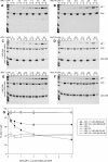
Both AIPL-1 and UNC-78 cooperate with the same ADF/cofilin isoform to promote actin filament disassembly in vitro. (A–F) Actin filament disassembly activity of GST-AIPL-1 and GST-UNC-78 were examined by F-actin sedimentation assays. F-actin (10 μM) was incubated with various concentrations (0–5 μM) of GST-AIPL-1 (A, C, E) or GST-UNC-78 (B, D, F) in the absence of ADF/cofilin (A, B) or in the presence of 20 μM UNC-60A (C, D) or 20 μM UNC-60B (E, F) for 30 min at room temperature. The mixtures were ultracentrifuged and fractionated into supernatants (s) and pellets (p) and analyzed by SDS–PAGE. Positions of bands of AIP1 (GST-AIPL-1 or GST-UNC-78), actin, UNC-60A, and UNC-60B are indicated on the right. (G) The results were quantitatively analyzed by densitometry. Percentages of actin in the pellets are plotted as a function of the concentrations of GST-AIPL-1 or GST-UNC-78. Data shown are mean ± SD of three independent experiments.
FIGURE 6:
FIGURE 6:
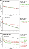
Both AIPL-1 and UNC-78 cooperate with the same ADF/cofilin isoform to promote the rate of actin filament disassembly in vitro. Kinetics of actin filament disassembly was analyzed by light scattering assays. F-actin was diluted to 4 μM in the absence of ADF/cofilin (A) or in the presence of 5 μM UNC-60A (B) or 5 μM UNC-60B (C). Experiments were performed in the absence of GST-AIPL-1 or GST-UNC-78 (black lines) or in the presence of GST-UNC-78 (green lines) or GST-AIPL-1 (red lines). Light scattering (arbitrary units) is plotted as a function of time (seconds).
FIGURE 7:
FIGURE 7:
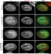
AIPL-1 cooperates with UNC-60B in organizing sarcomeric actin filaments in embryonic body wall muscle. Twofold-stage embryos of wild-type (A–C), unc-60B(r398) (D–F), aipl-1(ok1019) (G–I), and unc-60B(r398) aipl-1(ok1019) (J–L) worms were stained for actin (A, D, G, J) and UNC-60B (B, E, H, K). Merged images are shown in C, F, I, and L (actin in green and UNC-60B in red). Bar, 10 μm.
FIGURE 8:
FIGURE 8:
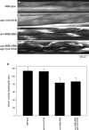
AIPL-1 is not required for organized sarcomeric actin assembly in adult body wall muscle. Adult wild-type (A), aipl-1(ok1019) (B), unc-60B(r398) (C), and unc-60B(r398) aipl-1(ok1019) (D) worms were stained for F-actin by tetramethylrhodamine-phalloidin. Bar, 20 μm. (E) Motility of wild-type, aipl-1(ok1019), unc-60B(r398), and unc-60B(r398) aipl-1(ok1019) worm. The aipl-1(ok1019) mutation did not significantly alter motility in wild-type or unc-60B(r398) background. Data are mean ± SD, n = 10.
FIGURE 9:
FIGURE 9:
AIPL-1 compensates for the function of UNC-78 in the body wall muscle. Adult worms of wild type (A–C), unc-78(gk27) (D–F), or unc-78(gk27) with a transgene for expression of GFP-AIPL-1 (G–I) were stained for F-actin by tetramethylrhodamine-phalloidin (A, D, G). Micrographs of the GFP fluorescence are shown in B, E, and H. Merged images are shown in C, F, and I (F-actin in red and GFP in green). Bar, 100 μm. Expression of GFP-AIPL-1 was forced in adult body wall muscle by the myo-3 promoter (Okkema et al., 1993). (J) Worm motility of wild type, unc-78(gk27), and unc-78(gk27) with a transgene for expression of GFP-AIPL-1 (three independent transgenic lines). Data are mean ± SD, n = 10. Motility of unc-78(gk27) with the transgene was significantly different (p < 0.001, asterisks) from that of unc-27(gk27) without the transgene.

Similar articles

Cited by

References

    1. Agrawal PB, et al. Nemaline myopathy with minicores caused by mutation of the CFL2 gene encoding the skeletal muscle actin-binding protein, cofilin-2. Am J Hum Genet. 2007;80:162–167. - PMC - PubMed
    1. Aizawa H, Katadae M, Maruya M, Sameshima M, Murakami-Murofushi K, Yahara I. Hyperosmotic stress-induced reorganization of actin bundles in Dictyostelium cells over-expressing cofilin. Genes Cells. 1999;4:311–324. - PubMed
    1. Allwood EG, Anthony RG, Smertenko AP, Reichelt S, Drobak BK, Doonan JH, Weeds AG, Hussey PJ. Regulation of the pollen-specific actin-depolymerizing factor LlADF1. Plant Cell. 2002;14:2915–2927. - PMC - PubMed
    1. Andrianantoandro E, Pollard TD. Mechanism of actin filament turnover by severing and nucleation at different concentrations of ADF/cofilin. Mol Cell. 2006;24:13–23. - PubMed
    1. Anyanful A, Ono K, Johnsen RC, Ly H, Jensen V, Baillie DL, Ono S. The RNA-binding protein SUP-12 controls muscle-specific splicing of the ADF/cofilin pre-mRNA in C. elegans. J Cell Biol. 2004;167:639–647. - PMC - PubMed

Publication types

MeSH terms

LinkOut - more resources