Review
. 2012 Apr;18(2):169-79.
doi: 10.1177/1073858411399925. Epub 2011 May 5.

In utero electroporation as a tool for genetic manipulation in vivo to study psychiatric disorders: from genes to circuits and behaviors

Affiliations
Review

In utero electroporation as a tool for genetic manipulation in vivo to study psychiatric disorders: from genes to circuits and behaviors

Yu Taniguchi et al. Neuroscientist. 2012 Apr.

Abstract

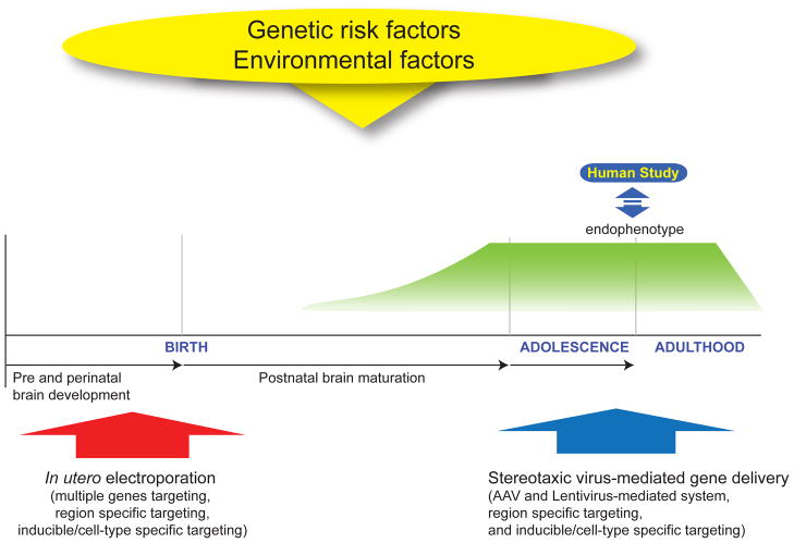
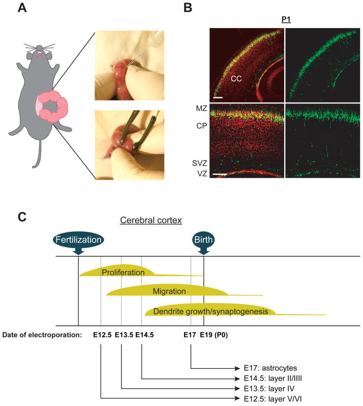
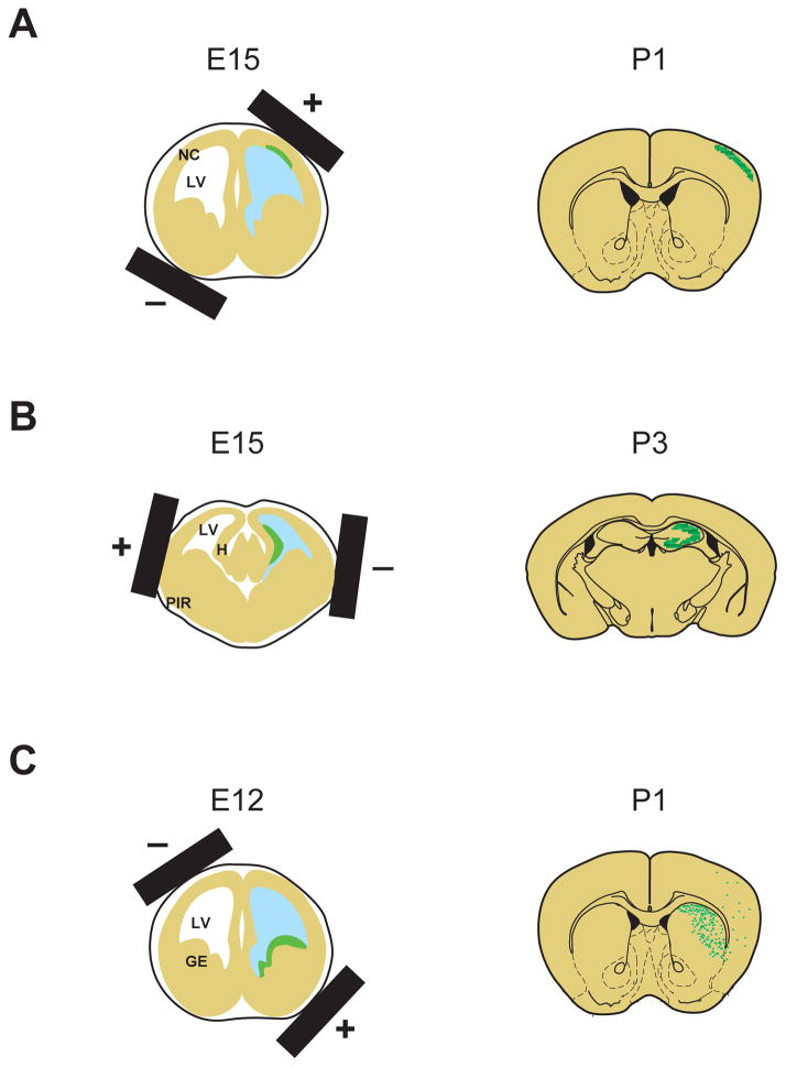
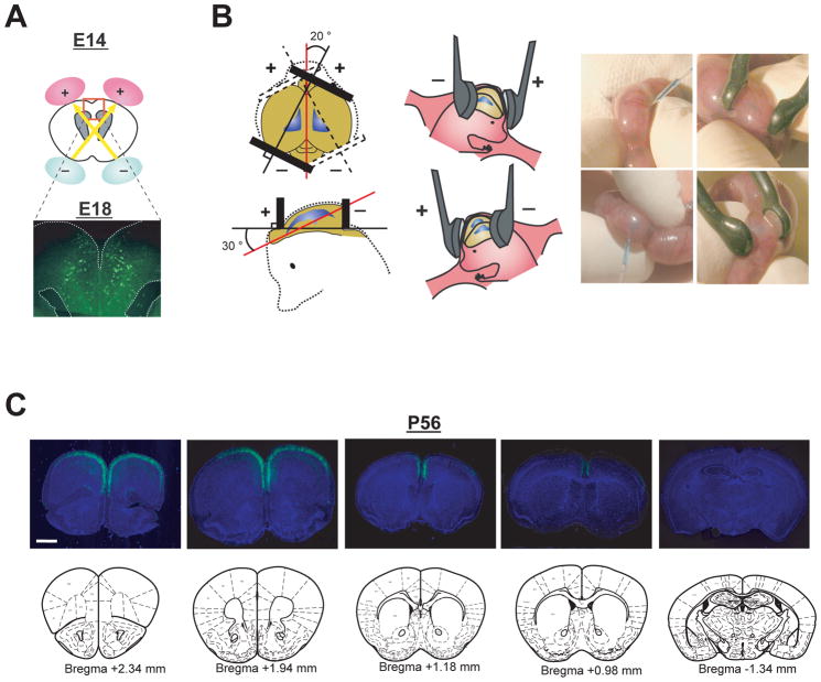
Many genetic risk factors for major mental disorders have key roles in brain development. Thus, exploring the roles for these genetic factors for brain development at the molecular, cellular, and neuronal circuit level is crucial for discovering how genetic disturbances affect high brain functions, which ultimately lead to disease pathologies. However, it is a tremendously difficult task, given that most mental disorders have genetic complexities in which many genetic risk factors have multiple roles in different cell types and brain regions over a time-course dependent manner. Furthermore, some genetic risk factors are likely to act epistatically in common molecular pathways. For this reason, a technique for spatial and temporal manipulation of multiple genes is necessary for understanding how genetic disturbances contribute to disease etiology. Here, the authors will review the said technique, in utero electroporation, which investigates the molecular disease pathways in rodent models for major mental disorders. This technique is also useful to examine the effect of genetic risks at the behavioral level. Furthermore, the authors will discuss the recent progress of this technology, such as inducible and cell type-specific targeting, as well as nonepisomal genetic manipulation, which provide further availability of this technique for research on major mental disorders.


Conflict of interest statement

Declaration of Conflicting Interests

The authors disclosed no conflicts of interests with respect to the authorship and/or publication of this article.

Figures

Figure 1
Figure 1. Animals models via Genetic manipulation in the developmental trajectory
Genetic manipulation techniques, such as in utero electroporation and stereotaxic virus-mediated gene delivery, are useful for examining the role for genetic risk factors for brain development and resultant higher brain function. The typical onset of psychiatric symptoms in most major mental disorders, such as schizophrenia and bipolar disorder, occurs during adolescence and young adulthood, whereas some psychiatric conditions, such as autism spectrum disorder, develop in childhood. Thus, it is important to understand the role for genetic risk factors in the developmental trajectory by segregating their functions at specific developmental stages in order to address these mechanisms precisely.
Figure 2
Figure 2. Genetic manipulation via in utero electroporation in selective cell population in the cerebral cortex
(A) Images displaying injection of DNA solution into lateral ventricle followed by gene delivery into the ventricular zone (VZ) via electroporation by holding the embryos with forceps-type electrodes. (B) Representative images of GFP-positive cells distributed at the cortical plate (CP) in the cerebral cortex at postnatal day 1 (P1); GFP expression constructs were introduced at embryonic day 15 (E15). CC, cerebral cortex; MZ, marginal zone; SVZ, subventricular zone; VZ, ventricular zone. Red, Nucleus. Scale bars, 100 μm. Adapted from the Kamiya (Kamiya 2009). (C) Schematic representation of the genetic manipulation in the selective cell population in the cerebral cortex. Electroporation directed into progenitor cells in the neocortical ventricular zone at E12.5, E13.5, E14.5, and E17 allows for the modulation of gene expression in a lineage of pyramidal neurons in the layer V/VI, layer IV, layer II/III, as well as astrocytes, respectively.
Figure 3
Figure 3. Region-specific gene targeting by in utero electroporation
Schematic representation of in utero electroporation for gene targeting into specific regions depending on the direction of electroporation. (A) For manipulating the cells in the neocortex, the neocortical neuroepithelium is targeted by placing the positive electrode on the dorsal lateral side. LV, lateral ventricle; NC, neocortex. (B) For manipulating the cells in the hippocampus, ammonic neuroepithelium is targeted via dorsal lateral placement of the positive electrode on the opposite side of their orientation for targeting the neocortex. PIR, piriform cortex; H, hippocampus. (C) For manipulating interneurons, ganglionic eminence, main sources of GABAergic interneurons, including cortical interneurons, are targeted by means of ventral lateral placement of the positive electrode at an approximate 30 degree outward angle from the horizontal plane. GE, ganglionic eminence.
Figure 4
Figure 4. Selective targeting to pyramidal neurons in the prefrontal cortex
(A) Schematic representation of bilateral in utero injection of constructs followed by their incorporation by electroporation into progenitor cells in the ventricular zone at E14. Migrating cells with GFP are visualized at E18 after injection of a GFP expression construct. (B) The embryo’s head in the uterus was held with a forceps-type electrode, consisting of two disc electrodes. The electrodes were oriented at approximately a 20 degree outward angle from the midline and a rough 30 degree angle downward from an imaginary line from the olfactory bulbs to the caudal side of cortical hemisphere. (C) Representative rostral-caudal image series of coronal sections of brains with GFP expression at P56 (+2.34 mm, +1.94 mm, +1.18 mm, +0.98 mm, and −1.34 mm from Bregma). Blue, nucleus. Scale bar, 1mm. Adapted from the Niwa et al (Niwa and others 2010).

Similar articles

Cited by

References

    1. Bai J, Ramos RL, Ackman JB, Thomas AM, Lee RV, LoTurco JJ. RNAi reveals doublecortin is required for radial migration in rat neocortex. Nat Neurosci. 2003;6(12):1277–83. - PubMed
    1. Bai J, Ramos RL, Paramasivam M, Siddiqi F, Ackman JB, LoTurco JJ. The role of DCX and LIS1 in migration through the lateral cortical stream of developing forebrain. Dev Neurosci. 2008;30(1–3):144–56. - PubMed
    1. Bill BR, Geschwind DH. Genetic advances in autism: heterogeneity and convergence on shared pathways. Curr Opin Genet Dev. 2009;19(3):271–8. - PMC - PubMed
    1. Borrell V, Yoshimura Y, Callaway EM. Targeted gene delivery to telencephalic inhibitory neurons by directional in utero electroporation. J Neurosci Methods. 2005;143(2):151–8. - PubMed
    1. Cetin A, Komai S, Eliava M, Seeburg PH, Osten P. Stereotaxic gene delivery in the rodent brain. Nat Protoc. 2006;1(6):3166–73. - PubMed

Publication types