. 2011 Jul 14;118(2):330-8.
doi: 10.1182/blood-2010-12-327353. Epub 2011 May 6.

Anti-inflammatory protein TSG-6 secreted by activated MSCs attenuates zymosan-induced mouse peritonitis by decreasing TLR2/NF-κB signaling in resident macrophages

Affiliations

Anti-inflammatory protein TSG-6 secreted by activated MSCs attenuates zymosan-induced mouse peritonitis by decreasing TLR2/NF-κB signaling in resident macrophages

Hosoon Choi et al. Blood. .

Abstract

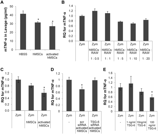
Human mesenchymal stem/progenitor cells (hMSCs) repair tissues and modulate immune systems but the mechanisms are not fully understood. We demonstrated that hMSCs are activated by inflammatory signals to secrete the anti-inflammatory protein, TNF-α-stimulated gene 6 protein (TSG-6) and thereby create a negative feedback loop that reduces inflammation in zymosan-induced peritonitis. The results demonstrate for the first time that TSG-6 interacts through the CD44 receptor on resident macrophages to decrease zymosan/TLR2-mediated nuclear translocation of the NF-κB. The negative feedback loop created by MSCs through TSG-6 attenuates the inflammatory cascade that is initiated by resident macrophages and then amplified by mesothelial cells and probably other cells of the peritoneum. Because inflammation underlies many pathologic processes, including immune responses, the results may explain the beneficial effects of MSCs and TSG-6 in several disease models.


Figures

Figure 1
Figure 1
Human MSCs and rhTSG-6 decreased the inflammatory response to zymosan-induced peritonitis. (A) Total cells in the lavage fluid. (B) Polymorphonuclear cells (PMN) in the lavage fluid. (C) Monocytes/macrophages in the lavage fluid. (D) Total cells in the lavage fluid harvested at 4 hours after administration of hMSCs, control hMSCs transfected with a scrambled siRNA (scr), hMSCs transfected with an siRNA for TSG-6, or rhTSG-6. Values are mean ± SD (n = 5 mice per time point; *P < .05; **P < .005; ***P < .0005).
Figure 2
Figure 2
Inhibition of zymosan-induced TNF-α expression in the peritoneum and in cultured macrophages. (A) ELISAs for mTNF-α in lavage fluid. Values are mean ± SD (n = 3; *P < .05). (B) Mouse-specific real-time RT-PCR assays for mTNF-α in macrophages stimulated with zymosan with or without various numbers of hMSCs. Data are expressed as mean and range of 2 values. (C) As in panel B except that the cocultures contained hMSCs (1:10 ratio to macrophages) and the hMSCs were either standard preparations or hMSCs activated to express TSG-6 by incubation with hTNF-α. (D) As in panel C except that the hMSCs were either activated hMSCs transfected with scrambled siRNA (scr) or activated hMSCs transfected with an siRNA for TSG-6. (E) As in panel D except murine macrophages cultured with zymosan with or without TSG-6. Values in panels C, D, and E are mean ± SD (n = 3; *P < .05).
Figure 3
Figure 3
TSG-6 inhibited nuclear translocation of NF-κB. Murine macrophages were incubated with zymosan with or without TSG-6. (A) Typical micrographs of immunocytochemistry are shown for cytoplasmic and nuclear distribution on NF-κB. (B) Quantification of data of micrographs from experiment in panel A. Values are mean ± SD for 3 random fields with at least 70 cells per field scored for each sample (n = 3; *P < .05; ***P < .0005).
Figure 4
Figure 4
Human MSCs and TSG-6 reduced amplification of the pro-inflammatory signals by mesothelial cells. (A) Human mesothelial (Mesoth) cells were incubated with zymosan and cultured alone or in cocultures with mouse macrophages (Mφ). Stimulation of the human mesothelial cells was assayed with human-specific RT-PCR assay for hIL-6. (B) As in panel A except TSG-6 was added to some of the cultures and stimulation of the human mesothelial cells was assayed with human-specific real-time RT-PCR assays for hIL-6, hIL-8, and hCCL2. (C) Systemic effects of the inflammatory cascade as indicated by plasma levels of mIL-6 assayed 8 hours after zymosan with or without subsequent infusion of hMSCs or TSG-6. Values are mean ± SD (n = 6 for HBSS, n = 3 for hMSCs and n = 4 for TSG-6; *P < .05; **P < .005; ***P < .005).
Figure 5
Figure 5
Three experimental strategies demonstrated CD44 was essential for inhibition by TSG-6. (A) Immunocytochemistry assay for expression of CD44 in clones of NF-κB reporter cells stably transfected with control plasmid (HEK-hTLR2-pcDNA) or plasmid containing complementary DNA for CD44 (HEK-hTLR2-CD44). (B) Assays for NF-κB signaling in the reporter cells expressing secreted alkaline phosphatase (SEAP). HEK-hTLR2-pcDNA and HEK-hTLR2-CD44 were incubated for 7 hours with zymosan with or without rhTSG-6. Values are mean ± SD (n = 3; **P < .001). (C) Murine macrophages pre-incubated for 15 minutes with control IgG (rat IgG2a) or blocking antibody for CD44 (CD44 KM81) and then incubated for 4 hours with zymosan and with or without activated hMSCs or TSG-6 (conditions as in Figure 2E). (D) Real-time RT-PCR assays with mouse-specific primers on resident macrophages isolated from wild-type mice (CD44+/+) and transgenic mice (CD44−/−) after injection of zymosan followed by injection of HBSS (n = 9 for CD44+/+ and n = 5 for CD44−/−), 1.6 × 10⁶ hMSCs (n = 5 for CD44+/+ and n = 3 for CD44−/−) or 30 μg of rhTSG-6 (n = 3 for CD44+/+ and CD44−/−). Values are mean ± SD (*P < .05; **P < .005).
Figure 6
Figure 6
Illustration of anti-inflammatory action of hMSCs mediated mainly through TSG-6. (1) Zymosan activated macrophages via TLR2. (2) Activated NF-κB increased the expression of pro-inflammatory cytokines. (3) HMSCs were activated by the pro-inflammatory cytokines to secrete TSG-6. (4) TSG-6 negatively regulated the TLR2-mediated responses through CD44.

References

    1. Friedenstein AJ, Gorskaja JF, Kulagina NN. Fibroblast precursors in normal and irradiated mouse hematopoietic organs. Exp Hematol. 1976;4(5):267–274. - PubMed
    1. Owen M, Friedenstein AJ. Stromal stem cells: marrow-derived osteogenic precursors. Ciba Found Symp. 1988;136:42–60. - PubMed
    1. Eaves CJ, Cashman JD, Sutherland HJ, et al. Molecular analysis of primitive hematopoietic cell proliferation control mechanisms. Ann N Y Acad Sci. 1991;628:298–306. - PubMed
    1. Caplan AI. Mesenchymal stem cells. J Orthop Res. 1991;9(5):641–650. - PubMed
    1. Dominici M, Le Blanc K, Mueller I, et al. Minimal criteria for defining multipotent mesenchymal stromal cells. The International Society for Cellular Therapy position statement. Cytotherapy. 2006;8(4):315–317. - PubMed

Publication types

MeSH terms