

Susceptible stages in Schwann cells for NF1-associated plexiform neurofibroma development
- PMID: 21551250
- PMCID: PMC3145496
- DOI: 10.1158/0008-5472.CAN-10-4577
Susceptible stages in Schwann cells for NF1-associated plexiform neurofibroma development
Abstract
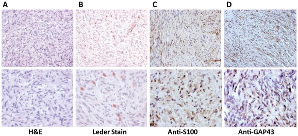
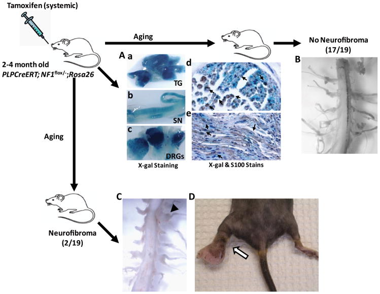
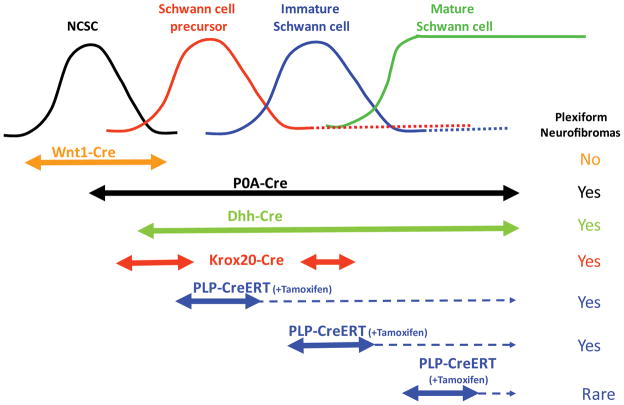
Stem cells are under strict regulation by both intrinsic factors and the microenvironment. There is increasing evidence that many cancers initiate through acquisition of genetic mutations (loss of intrinsic control) in stem cells or their progenitors, followed by alterations of the surrounding microenvironment (loss of extrinsic control). In neurofibromatosis type 1 (NF1), deregulation of Ras signaling results in development of multiple neurofibromas, complex tumors of the peripheral nerves. Neurofibromas arise from the Schwann cell lineage following loss of function at the NF1 locus, which initiates a cascade of interactions with other cell types in the microenvironment and additional cell autonomous modifications. In this study, we sought to identify whether a temporal "window of opportunity" exists during which cells of the Schwann cell lineage can give rise to neurofibromas following loss of NF1. We showed that acute loss of NF1 in both embryonic and adult Schwann cells can lead to neurofibroma formation. However, the embryonic period when Schwann cell precursors and immature Schwann cells are most abundant coincides with enhanced susceptibility to plexiform neurofibroma tumorigenesis. This model has important implications for understanding early cellular events that dictate neurofibroma development, as well as for the development of novel therapies targeting these tumors.
©2011 AACR.
Figures
Similar articles
-
The role of nerve microenvironment for neurofibroma development.Oncotarget. 2016 Sep 20;7(38):61500-61508. doi: 10.18632/oncotarget.11133. Oncotarget. 2016. PMID: 27517146 Free PMC article.
-
Perinatal or adult Nf1 inactivation using tamoxifen-inducible PlpCre each cause neurofibroma formation.Cancer Res. 2011 Jul 1;71(13):4675-85. doi: 10.1158/0008-5472.CAN-10-4558. Epub 2011 May 6. Cancer Res. 2011. PMID: 21551249 Free PMC article.
-
Plexiform and dermal neurofibromas and pigmentation are caused by Nf1 loss in desert hedgehog-expressing cells.Cancer Cell. 2008 Feb;13(2):105-16. doi: 10.1016/j.ccr.2007.12.027. Cancer Cell. 2008. PMID: 18242511 Free PMC article.
-
Genetically engineered mouse models shed new light on the pathogenesis of neurofibromatosis type I-related neoplasms of the peripheral nervous system.Brain Res Bull. 2012 May 1;88(1):58-71. doi: 10.1016/j.brainresbull.2011.08.005. Epub 2011 Aug 10. Brain Res Bull. 2012. PMID: 21855613 Free PMC article. Review.
-
Plexiform neurofibroma genesis: questions of Nf1 gene dose and hyperactive mast cells.Curr Opin Hematol. 2010 Jul;17(4):287-93. doi: 10.1097/MOH.0b013e328339511b. Curr Opin Hematol. 2010. PMID: 20571392 Free PMC article. Review.
Cited by
-
New insights into the neurofibroma tumor cells of origin.Neurooncol Adv. 2019 Nov 10;2(Suppl 1):i13-i22. doi: 10.1093/noajnl/vdz044. eCollection 2020 Jul. Neurooncol Adv. 2019. PMID: 32642729 Free PMC article. Review.
-
[Gene therapy strategies and prospects for neurofibromatosis type 1].Zhongguo Xiu Fu Chong Jian Wai Ke Za Zhi. 2024 Jan 15;38(1):1-8. doi: 10.7507/1002-1892.202309071. Zhongguo Xiu Fu Chong Jian Wai Ke Za Zhi. 2024. PMID: 38225833 Free PMC article. Chinese.
-
Stem-like cells drive NF1-associated MPNST functional heterogeneity and tumor progression.Cell Stem Cell. 2021 Aug 5;28(8):1397-1410.e4. doi: 10.1016/j.stem.2021.04.029. Epub 2021 May 18. Cell Stem Cell. 2021. PMID: 34010628 Free PMC article.
-
Neurofibroma Development in Neurofibromatosis Type 1: Insights from Cellular Origin and Schwann Cell Lineage Development.Cancers (Basel). 2022 Sep 17;14(18):4513. doi: 10.3390/cancers14184513. Cancers (Basel). 2022. PMID: 36139671 Free PMC article. Review.
-
Cells of origin in the embryonic nerve roots for NF1-associated plexiform neurofibroma.Cancer Cell. 2014 Nov 10;26(5):695-706. doi: 10.1016/j.ccell.2014.09.009. Epub 2014 Oct 30. Cancer Cell. 2014. PMID: 25446898 Free PMC article.
References
-
- Reya T, Morrison SJ, Clarke MF, Weissman IL. Stem cells, cancer, and cancer stem cells. Nature. 2001;414:105–11. - PubMed
-
- Singh SK, Hawkins C, Clarke ID, et al. Identification of human brain tumour initiating cells. Nature. 2004;432:396–401. - PubMed
-
- Barker N, Ridgway RA, van Es JH, et al. Crypt stem cells as the cells-of-origin of intestinal cancer. Nature. 2009;457:608–11. - PubMed
Publication types
MeSH terms
Substances
Grants and funding
LinkOut - more resources
Full Text Sources
Molecular Biology Databases
Research Materials
Miscellaneous
