. 2011 Jul;10(7):1137-48.
doi: 10.1158/1535-7163.MCT-10-1028. Epub 2011 May 6.

Expression signatures of the lipid-based Akt inhibitors phosphatidylinositol ether lipid analogues in NSCLC cells

Affiliations

Expression signatures of the lipid-based Akt inhibitors phosphatidylinositol ether lipid analogues in NSCLC cells

Chunyu Zhang et al. Mol Cancer Ther. 2011 Jul.

Abstract

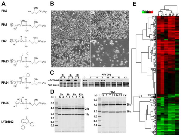
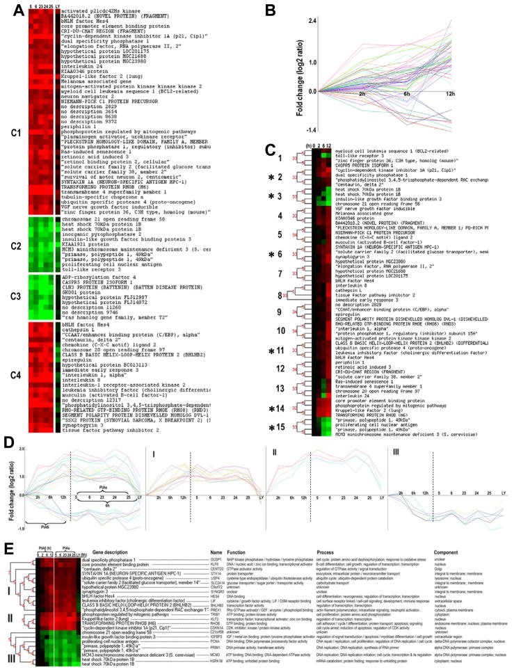
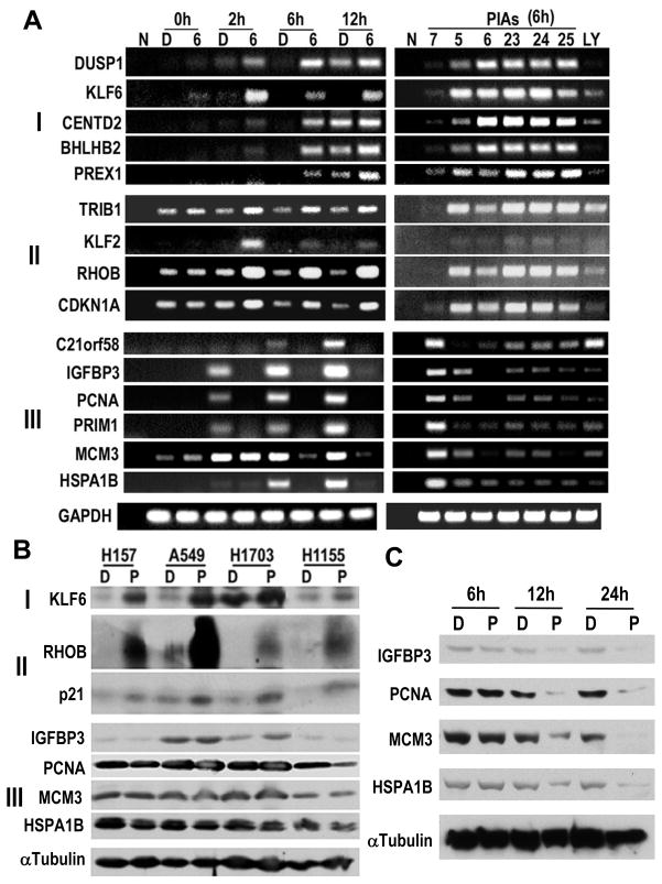
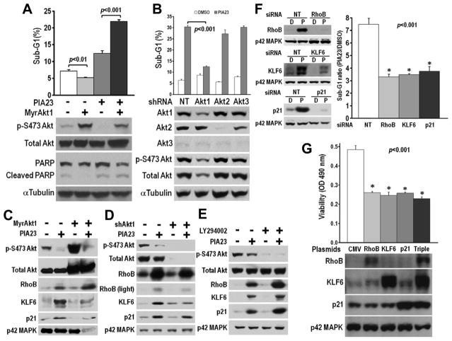
Activation of the serine/threonine kinase Akt contributes to the formation, maintenance, and therapeutic resistance of cancer, which is driving development of compounds that inhibit Akt. Phosphatidylinositol ether lipid analogues (PIA) are analogues of the products of phosphoinositide-3-kinase (PI3K) that inhibit Akt activation, translocation, and the proliferation of a broad spectrum of cancer cell types. To gain insight into the mechanism of PIAs, time-dependent transcriptional profiling of five active PIAs and the PI3K inhibitor LY294002 (LY) was conducted in non-small cell lung carcinoma cells using high-density oligonucleotide arrays. Gene ontology analysis revealed that genes involved in apoptosis, wounding response, and angiogenesis were upregulated by PIAs, whereas genes involved in DNA replication, repair, and mitosis were suppressed. Genes that exhibited early differential expression were partitioned into three groups; those induced by PIAs only (DUSP1, KLF6, CENTD2, BHLHB2, and PREX1), those commonly induced by PIAs and LY (TRIB1, KLF2, RHOB, and CDKN1A), and those commonly suppressed by PIAs and LY (IGFBP3, PCNA, PRIM1, MCM3, and HSPA1B). Increased expression of the tumor suppressors RHOB (RhoB), KLF6 (COPEB), and CDKN1A (p21Cip1/Waf1) was validated as an Akt-independent effect that contributed to PIA-induced cytotoxicity. Despite some overlap with LY, active PIAs have a distinct expression signature that contributes to their enhanced cytotoxicity.


Conflict of interest statement

The authors have no conflict of interest.

Figures

Figure 1
Figure 1
Optimization of PIA treatment and oligonucleotide microarray analysis. A) The chemical structures of the inactive PIA7, 5 active PIAs and PI3K inhibitor LY294002. B) Cellular morphological alterations induced by PIA treatment. H157 cells were incubated with 10 μM PIA6 dissolved in DMSO in RPMI1640 + 0.1% FBS media for the indicated times. C) Evaluation of Akt inhibition in samples collected for microarray analysis. Parallel H157 cell samples were collected for analysis of p-S473 Akt by immunoblot, alongside microarray samples for RNA extraction, in time course and PIA comparison experiments as described in Materials and Methods. D = DMSO, 5, 6, 7, 23, 24, 25 = 10 μM PIA-treated samples, LY = 10 μM LY294002-treated sample. D) Assessment of RNA quality and integrity in microarray samples using the Bioanalyzer Nanochip. Prominent bands indicate positions of 28s and 18s rRNA. E) Clustered heat map showing PIA-altered genes. Red color = induction, green color = suppression, black color = no change in expression. Complete linkage hierarchical clustering of PIA-regulated gene expression changes in H157 cells was performed with uncentered correlation as described in Materials and Methods.
Figure 2
Figure 2
Identification of genes that changed early and in common with PIA or LY treatment. A) Filtered set of common PIA-regulated genes grouped using k-Means clustering. C1 = genes that were induced by all 5 active PIAs and the PI3K inhibitor LY, C2 = genes suppressed by active PIAs and LY, C3 = genes suppressed by PIAs but not LY, C4 = genes that were induced by PIAs only, not LY. B) Temporal dynamics of common PIA-regulated genes. Expression levels of common PIA-regulated genes from figure 2A were extracted from PIA6 time course experiment and are depicted at the different time points. C) 15 temporal clusters of genes from figure 2B whose expression changed in a similar manner generated by CAGED program. The asterisks mark clusters (2, 3, 6, 11, 14 and 15) that were chosen for further analysis. D) Depiction of genes that were commonly regulated by PIAs and their temporal dynamics. Group I = genes induced early by PIAs but not LY, group II = genes induced early and in common with LY, group III = genes suppressed early by PIAs and in common with LY. E) Identity, function and cellular location of Group I-III genes.
Figure 3
Figure 3
Validation of microarray data by RT-PCR and immunoblotting. A) Validation of oligonucleotide microarray data by semi-quantitative RT-PCR. Expression of genes from groups I, II and III were evaluated in time course (left panels) and PIA comparison experiments (right panels). N = RNA omitted negative control. B) Immunoblotting analysis of KLF6, RhoB and p21 protein induction in vitro by PIA23 (6h) in 4 NSCLC cell lines. C) Protein expression of PIA-repressed transcripts in H157 cells decreases at later time points. D = DMSO, P = PIA23 (10 μM).
Figure 4
Figure 4
Relevance of active Akt and induced genes to PIA-induced cytotoxicity. A) FACS analysis of sub-G1 DNA content and immunoblotting analysis of active Akt and cleaved PARP in MyrAkt1-expressing H157 cells treated with PIA23 (18h). B) Validation of Akt isoform specific knockdown by shAkt1, 2 or 3 in A549 cells and effect of individual stable knockdown on expression level of total and active Akt, as well as PIA23-induced apoptosis (24h). C) Expression of RhoB, KLF6 and p21 in MyrAkt1 or vector-expressing H157 cells treated with PIA23 (18h). D) Expression of RhoB, KLF6 and p21 in shAkt1 or non-targeting vector transfected A549 cells treated with PIA23 (24h). E) Expression of RhoB, KLF6 and p21 in H157 cells pretreated with LY294002 (0.5h), followed by treatment with PIA23 (6h). F) Effect of transient RhoB, KLF6, p21 or non-targeting (NT) siRNA transfection (48h) on protein induction by PIA23 (6h) and PIA-induced sub-G1 content in H157 cells (12h). G) Effect of overexpression of RhoB, KLF6 and p21 or the combination of all 3 genes on viability in H157 cells (48h). PIA23 and LY294002 = 10 μM. Data are means ± s.d. of triplicates and representative of 3 independent experiments.

References

    1. LoPiccolo J, Granville CA, Gills JJ, Dennis PA. Targeting Akt in cancer therapy. Anticancer Drugs. 2007;18:861–74. - PubMed
    1. Brognard J, Clark AS, Ni Y, Dennis PA. Akt/protein kinase B is constitutively active in non-small cell lung cancer cells and promotes cellular survival and resistance to chemotherapy and radiation. Cancer Res. 2001;61:3986–97. - PubMed
    1. Clark AS, West K, Streicher S, Dennis PA. Constitutive and inducible Akt activity promotes resistance to chemotherapy, trastuzumab, or tamoxifen in breast cancer cells. Mol Cancer Ther. 2002;1:707–17. - PubMed
    1. Granville CA, Memmott RM, Gills JJ, Dennis PA. Handicapping the race to develop inhibitors of the phosphoinositide 3-kinase/Akt/mammalian target of rapamycin pathway. Clin Cancer Res. 2006;12:679–89. - PubMed
    1. LoPiccolo J, Blumenthal GM, Bernstein WB, Dennis PA. Targeting the PI3K/Akt/mTOR pathway: effective combinations and clinical considerations. Drug Resist Updat. 2008;11:32–50. - PMC - PubMed

Publication types

MeSH terms