

Catalase overexpression in aortic smooth muscle prevents pathological mechanical changes underlying abdominal aortic aneurysm formation
- PMID: 21551275
- PMCID: PMC3154675
- DOI: 10.1152/ajpheart.00040.2011
Catalase overexpression in aortic smooth muscle prevents pathological mechanical changes underlying abdominal aortic aneurysm formation
Abstract
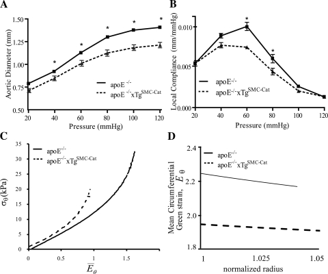
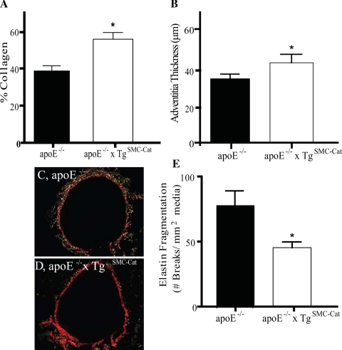
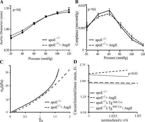
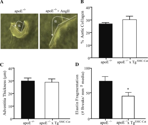
The causality of the associations between cellular and mechanical mechanisms of abdominal aortic aneurysm (AAA) formation has not been completely defined. Because reactive oxygen species are established mediators of AAA growth and remodeling, our objective was to investigate oxidative stress-induced alterations in aortic biomechanics and microstructure during subclinical AAA development. We investigated the mechanisms of AAA in an angiotensin II (ANG II) infusion model of AAA in apolipoprotein E-deficient (apoE(-/-)) mice that overexpress catalase in vascular smooth muscle cells (apoE(-/-)xTg(SMC-Cat)). At baseline, aortas from apoE(-/-)xTg(SMC-Cat) exhibited increased stiffness and the microstructure was characterized by 50% more collagen content and less elastin fragmentation. ANG II treatment for 7 days in apoE(-/-) mice altered the transmural distribution of suprarenal aortic circumferential strain (quantified by opening angle, which increased from 130 ± 1° at baseline to 198 ± 8° after 7 days of ANG II treatment) without obvious changes in the aortic microstructure. No differences in aortic mechanical behavior or suprarenal opening angle were observed in apoE(-/-)xTg(SMC-Cat) after 7 days of ANG II treatment. These data suggest that at the earliest stages of AAA development H(2)O(2) is functionally important and is involved in the control of local variations in remodeling across the vessel wall. They further suggest that reduced elastin integrity at baseline may predispose the abdominal aorta to aneurysmal mechanical remodeling.
Figures
Similar articles
-
Deficiency of cathepsin S attenuates angiotensin II-induced abdominal aortic aneurysm formation in apolipoprotein E-deficient mice.Cardiovasc Res. 2012 Dec 1;96(3):401-10. doi: 10.1093/cvr/cvs263. Epub 2012 Aug 7. Cardiovasc Res. 2012. PMID: 22871592 Free PMC article.
-
The Paraoxonase Gene Cluster Protects Against Abdominal Aortic Aneurysm Formation.Arterioscler Thromb Vasc Biol. 2017 Feb;37(2):291-300. doi: 10.1161/ATVBAHA.116.308684. Epub 2016 Dec 1. Arterioscler Thromb Vasc Biol. 2017. PMID: 27908891
-
Overexpression of catalase in vascular smooth muscle cells prevents the formation of abdominal aortic aneurysms.Arterioscler Thromb Vasc Biol. 2013 Oct;33(10):2389-96. doi: 10.1161/ATVBAHA.113.302175. Epub 2013 Aug 15. Arterioscler Thromb Vasc Biol. 2013. PMID: 23950141 Free PMC article.
-
Translational Relevance and Recent Advances of Animal Models of Abdominal Aortic Aneurysm.Arterioscler Thromb Vasc Biol. 2017 Mar;37(3):401-410. doi: 10.1161/ATVBAHA.116.308534. Epub 2017 Jan 5. Arterioscler Thromb Vasc Biol. 2017. PMID: 28062500 Review.
-
Three-dimensional microstructural changes in murine abdominal aortic aneurysms quantified using immunofluorescent array tomography.J Histochem Cytochem. 2012 Feb;60(2):97-109. doi: 10.1369/0022155411433066. Epub 2011 Dec 1. J Histochem Cytochem. 2012. PMID: 22140132 Free PMC article. Review.
Cited by
-
Hydrogen Sulfide Attenuates Aortic Remodeling in Aortic Dissection Associating with Moderated Inflammation and Oxidative Stress through a NO-Dependent Pathway.Antioxidants (Basel). 2021 Apr 27;10(5):682. doi: 10.3390/antiox10050682. Antioxidants (Basel). 2021. PMID: 33925479 Free PMC article.
-
Nanotherapies for Treatment of Cardiovascular Disease: A Case for Antioxidant Targeted Delivery.Curr Pathobiol Rep. 2019 Sep;7(3):47-60. doi: 10.1007/s40139-019-00196-4. Epub 2019 Jun 27. Curr Pathobiol Rep. 2019. PMID: 31396435 Free PMC article.
-
The role of phosphoinositide 3-kinases in immune-inflammatory responses: potential therapeutic targets for abdominal aortic aneurysm.Cell Cycle. 2022 Nov;21(22):2339-2364. doi: 10.1080/15384101.2022.2094577. Epub 2022 Jul 6. Cell Cycle. 2022. PMID: 35792922 Free PMC article. Review.
-
Caveolin 1 is critical for abdominal aortic aneurysm formation induced by angiotensin II and inhibition of lysyl oxidase.Clin Sci (Lond). 2014 Jun;126(11):785-94. doi: 10.1042/CS20130660. Clin Sci (Lond). 2014. PMID: 24329494 Free PMC article.
-
Oxidative Stress and the Pathogenesis of Aortic Aneurysms.Biomedicines. 2023 Dec 19;12(1):3. doi: 10.3390/biomedicines12010003. Biomedicines. 2023. PMID: 38275364 Free PMC article. Review.
References
-
- Anidjar S, Salzmann J, Gentric D, Lagneau P, Camilleri J, Michel J. Elastase-induced experimental aneurysms in rats. Circulation 82: 973–981, 1990 - PubMed
-
- Burke JM, Balian G, Ross R, Bornstein P. Synthesis of types I and III procollagen and collagen by monkey aortic smooth muscle cells in vitro. Biochemistry 16: 3243–3249, 1977 - PubMed
-
- Chatelain RE, D BN. Increased DNA replication in the arterial adventitia after aortic ligation. Hypertension 11: I130–I134, 1988 - PubMed
-
- Chiquet M, Renedo AS, Huber F, Flück M. How do fibroblasts translate mechanical signals into changes in extracellular matrix production? Matrix Biol 22: 73–80, 2003 - PubMed
Publication types
MeSH terms
Substances
Grants and funding
LinkOut - more resources
Full Text Sources
Molecular Biology Databases
Miscellaneous