

Doxycycline, a matrix metalloprotease inhibitor, reduces vascular remodeling and damage after cerebral ischemia in stroke-prone spontaneously hypertensive rats
- PMID: 21551278
- PMCID: PMC3129920
- DOI: 10.1152/ajpheart.01206.2010
Doxycycline, a matrix metalloprotease inhibitor, reduces vascular remodeling and damage after cerebral ischemia in stroke-prone spontaneously hypertensive rats
Abstract
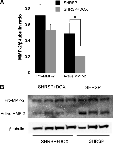
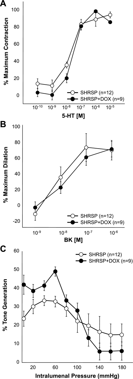
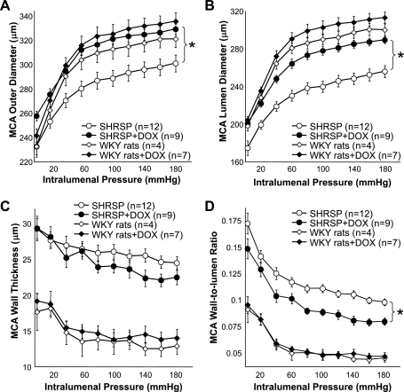
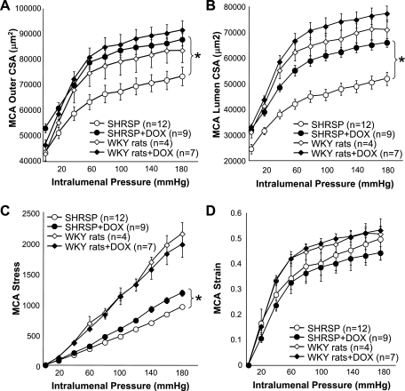
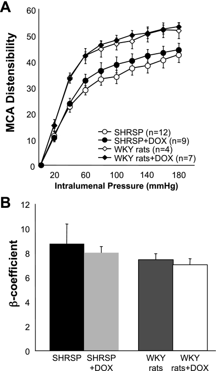
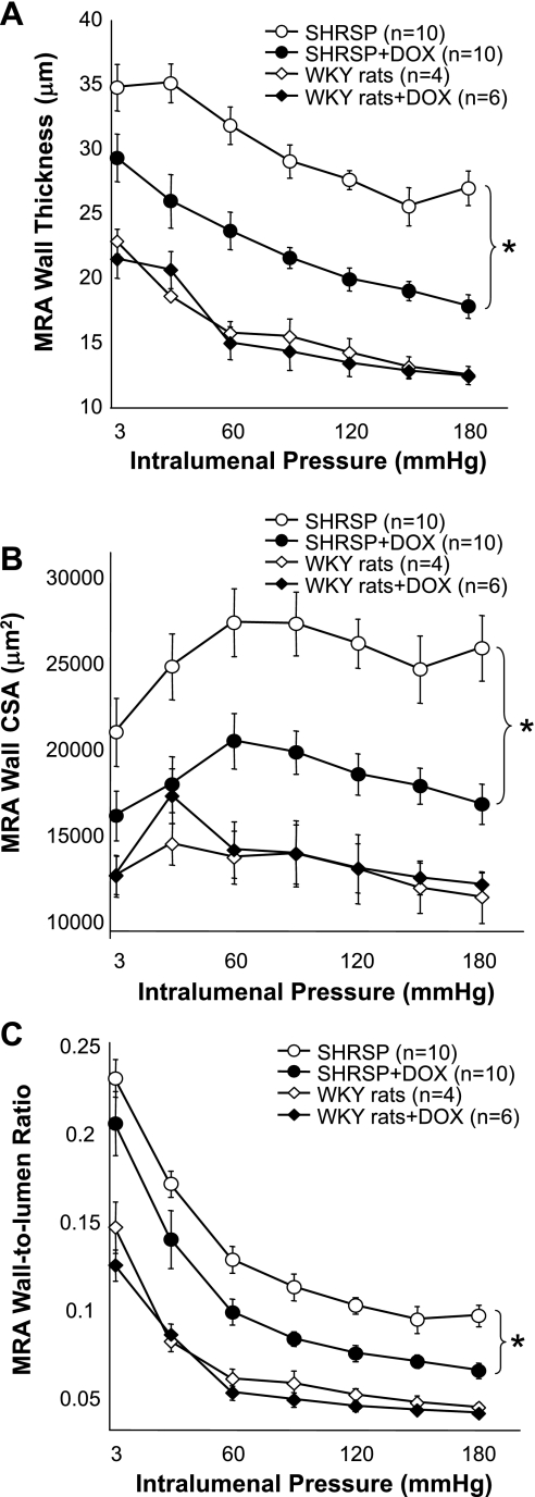
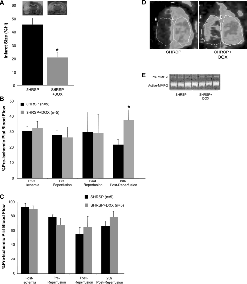
Matrix metalloproteases (MMPs) are a family of zinc peptidases involved in extracellular matrix turnover. There is evidence that increased MMP activity is involved in remodeling of resistance vessels in chronic hypertension. Thus we hypothesized that inhibition of MMP activity with doxycycline (DOX) would attenuate vascular remodeling. Six-week-old male stroke-prone spontaneously hypertensive rats (SHRSP) were treated with DOX (50 mg·kg(-1)·day(-1) in the drinking water) for 6 wk. Untreated SHRSP were controls. Blood pressure was measured by telemetry during the last week. Middle cerebral artery (MCA) and mesenteric resistance artery (MRA) passive structures were assessed by pressure myography. MMP-2 expression in aortas was measured by Western blot. All results are means ± SE. DOX caused a small increase in mean arterial pressure (SHRSP, 154 ± 1; SHRSP + DOX, 159 ± 3 mmHg; P < 0.001). Active MMP-2 expression was reduced in aorta from SHRSP + DOX (0.21 ± 0.06 vs. 0.49 ± 0.13 arbitrary units; P < 0.05). In the MCA, at 80 mmHg, DOX treatment increased the lumen (273.2 ± 4.7 vs. 238.3 ± 6.3 μm; P < 0.05) and the outer diameter (321 ± 5.3 vs. 290 ± 7.6 μm; P < 0.05) and reduced the wall-to-lumen ratio (0.09 ± 0.002 vs. 0.11 ± 0.003; P < 0.05). Damage after transient cerebral ischemia (transient MCA occlusion) was reduced in SHRSP + DOX (20.7 ± 4 vs. 45.5 ± 5% of hemisphere infarcted; P < 0.05). In the MRA, at 90 mmHg DOX, reduced wall thickness (29 ± 1 vs. 22 ± 1 μm; P < 0.001) and wall-to-lumen ratio (0.08 ± 0.004 vs. 0.11 ± 0.008; P < 0.05) without changing lumen diameter. These results suggest that MMPs are involved in hypertensive vascular remodeling in both the peripheral and cerebral vasculature and that DOX reduced brain damage after cerebral ischemia.
Figures
Similar articles
-
Tumor necrosis factor-α inhibition attenuates middle cerebral artery remodeling but increases cerebral ischemic damage in hypertensive rats.Am J Physiol Heart Circ Physiol. 2014 Sep 1;307(5):H658-69. doi: 10.1152/ajpheart.00018.2014. Epub 2014 Jul 11. Am J Physiol Heart Circ Physiol. 2014. PMID: 25015967 Free PMC article.
-
Spironolactone improves structure and increases tone in the cerebral vasculature of male spontaneously hypertensive stroke-prone rats.Microvasc Res. 2007 May;73(3):198-205. doi: 10.1016/j.mvr.2006.12.001. Epub 2007 Jan 23. Microvasc Res. 2007. PMID: 17250855 Free PMC article.
-
Effects of spironolactone on cerebral vessel structure in rats with sustained hypertension.Am J Hypertens. 2011 Jun;24(6):708-15. doi: 10.1038/ajh.2011.20. Epub 2011 Feb 24. Am J Hypertens. 2011. PMID: 21350432 Free PMC article.
-
Focal Ischemic Injury with Complex Middle Cerebral Artery in Stroke-Prone Spontaneously Hypertensive Rats with Loss-Of-Function in NADPH Oxidases.PLoS One. 2015 Sep 21;10(9):e0138551. doi: 10.1371/journal.pone.0138551. eCollection 2015. PLoS One. 2015. PMID: 26389812 Free PMC article.
-
The effects of hypertension on the cerebral circulation.Am J Physiol Heart Circ Physiol. 2013 Jun 15;304(12):H1598-614. doi: 10.1152/ajpheart.00490.2012. Epub 2013 Apr 12. Am J Physiol Heart Circ Physiol. 2013. PMID: 23585139 Free PMC article. Review.
Cited by
-
Regulation of myogenic tone and structure of parenchymal arterioles by hypertension and the mineralocorticoid receptor.Am J Physiol Heart Circ Physiol. 2015 Jul 1;309(1):H127-36. doi: 10.1152/ajpheart.00168.2015. Epub 2015 Apr 24. Am J Physiol Heart Circ Physiol. 2015. PMID: 25910805 Free PMC article.
-
Is there new hope for therapeutic matrix metalloproteinase inhibition?Nat Rev Drug Discov. 2014 Dec;13(12):904-27. doi: 10.1038/nrd4390. Epub 2014 Nov 7. Nat Rev Drug Discov. 2014. PMID: 25376097 Review.
-
Matrix metalloproteinases promote arterial remodeling in aging, hypertension, and atherosclerosis.Hypertension. 2015 Apr;65(4):698-703. doi: 10.1161/HYPERTENSIONAHA.114.03618. Epub 2015 Feb 9. Hypertension. 2015. PMID: 25667214 Free PMC article. Review. No abstract available.
-
Long-term use of doxycycline can improve chronic asthma and possibly remodeling: the result of a pilot observation.J Asthma Allergy. 2012;5:33-7. doi: 10.2147/JAA.S31402. Epub 2012 Aug 8. J Asthma Allergy. 2012. PMID: 22923997 Free PMC article.
-
Tumor necrosis factor-α inhibition attenuates middle cerebral artery remodeling but increases cerebral ischemic damage in hypertensive rats.Am J Physiol Heart Circ Physiol. 2014 Sep 1;307(5):H658-69. doi: 10.1152/ajpheart.00018.2014. Epub 2014 Jul 11. Am J Physiol Heart Circ Physiol. 2014. PMID: 25015967 Free PMC article.
References
-
- Bassiouny HS, Song RH, Hong XF, Singh A, Kocharyan H, Glagov S. Flow regulation of 72-kD collagenase IV (MMP-2) after experimental arterial injury. Circulation 98: 157–163, 1998 - PubMed
-
- Baumbach GL, Ghoneim S. Vascular remodeling in hypertension. Scanning Microsc 7: 137–142, 1993 - PubMed
-
- Baumbach GL, Hajdu MA. Mechanics and composition of cerebral arterioles in renal and spontaneously hypertensive rats. Hypertension 21: 816–826, 1993 - PubMed
-
- Castro MM, Rizzi E, Figueiredo-Lopes L, Fernandes K, Bendhack LM, Pitol DL, Gerlach RF, Tanus-Santos JE. Metalloproteinase inhibition ameliorates hypertension and prevents vascular dysfunction and remodeling in renovascular hypertensive rats. Atherosclerosis 198: 320–331, 2008 - PubMed
-
- Castro MM, Rizzi E, Prado CM, Rossi MA, Tanus-Santos JE, Gerlach RF. Imbalance between matrix metalloproteinases and tissue inhibitor of metalloproteinases in hypertensive vascular remodeling. Matrix Biol 29: 194–201, 2010 - PubMed
Publication types
MeSH terms
Substances
Grants and funding
LinkOut - more resources
Full Text Sources
Medical
Miscellaneous