. 2011 Jul;90(7):894-9.
doi: 10.1177/0022034511407918. Epub 2011 May 6.

Bisphosphonates inhibit expression of p63 by oral keratinocytes

Affiliations

Bisphosphonates inhibit expression of p63 by oral keratinocytes

E L Scheller et al. J Dent Res. 2011 Jul.

Abstract

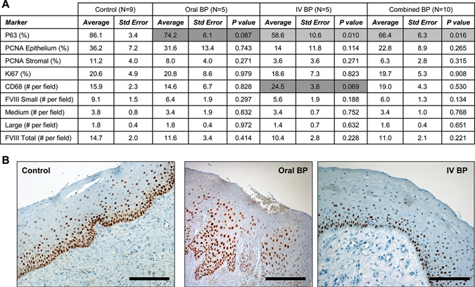
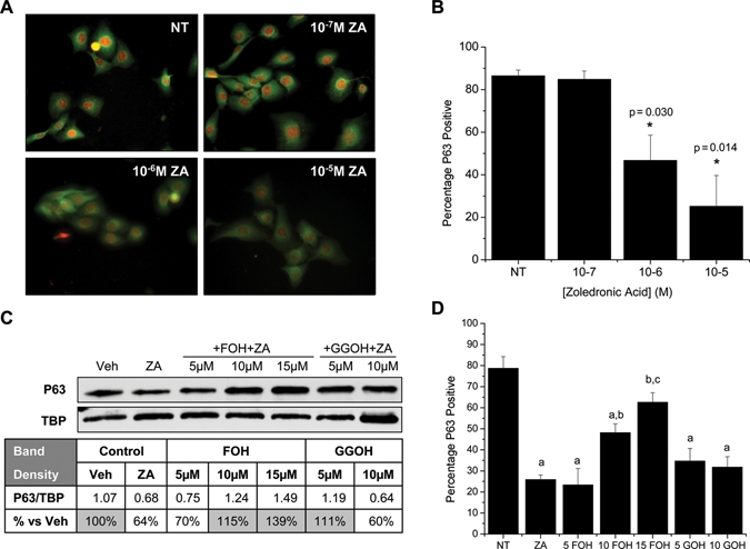
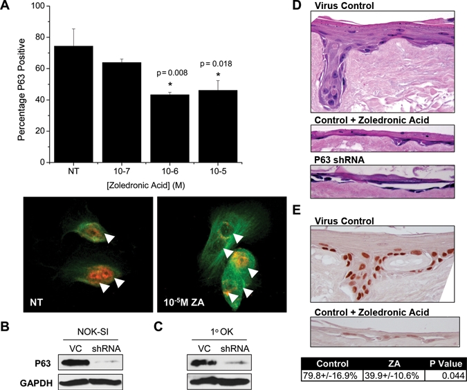
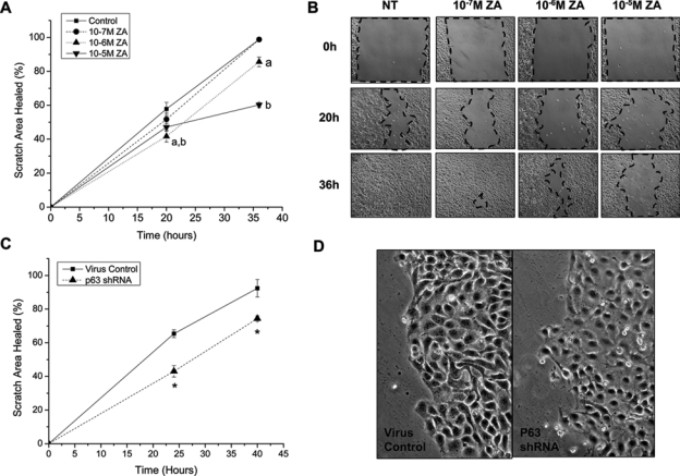
Osteonecrosis of the jaw (ONJ), a side-effect of bisphosphonate therapy, is characterized by exposed bone that fails to heal within eight weeks. Healing time of oral epithelial wounds is decreased in the presence of amino-bisphosphonates; however, the mechanism remains unknown. We examined human tissue from individuals with ONJ and non-bisphosphonate-treated control individuals to identify changes in oral epithelium and connective tissue. Oral and intravenous bisphosphonate-treated ONJ sites had reduced numbers of basal epithelial progenitor cells, as demonstrated by a 13.8±1.1% and 31.9±5.8% reduction of p63 expression, respectively. No significant differences in proliferation rates, vessel density, or macrophage number were noted. In vitro treatment of clonal and primary oral keratinocytes with zoledronic acid (ZA) inhibited p63, and expression was rescued by the addition of mevalonate pathway intermediates. In addition, both ZA treatment and p63 shRNA knock-down impaired formation of 3D Ex Vivo Produced Oral Mucosa Equivalents (EVPOME) and closure of an in vitro scratch assay. Analysis of our data suggests that bisphosphonate treatment may delay oral epithelial healing by interfering with p63-positive progenitor cells in the basal layer of the oral epithelium in a mevalonate-pathway-dependent manner. This delay in healing may increase the likelihood of osteonecrosis developing in already-compromised bone.


Conflict of interest statement

The authors declare no potential conflicts of interest with respect to the authorship and/or publication of this article.

Figures

Figure 1.
Figure 1.
Bisphosphonates inhibit expression of p63 in basal oral keratinocytes. (A) Tabulated immunohistochemistry results. (B) Representative images of control, Oral-BP, and IV-BP epithelial staining for p63. Results are reported as average ± standard error of the mean.
Figure 2.
Figure 2.
Zoledronic acid inhibits p63 in clonal oral keratinocytes. (A) Immunocytochemistry depicting p63 nuclear stain (red) and B-tubulin cytoplasmic stain (green). (B) Percentage p63-positive cells as calculated by applying a common threshold to matched p63-stained images and comparison with total number of cells per field. Results are reported as the average of 3 independent experiments (N = 3), with 3 random fields analyzed per treatment regimen per experiment. (C) Representative Western blot of nuclear extracts from NOK-SI treated with 10-5 M ZA, vehicle control, FOH, or GGOH. Band densitometry was used to normalize p63 to the TATA binding protein (TBP) loading standard. (D) Percentage p63-positive cells calculated as in (B) after treatment with vehicle (NT), 10-5 M ZA, ZA+5-15 µM FOH, or ZA+5-10 µM GGOH. (a) Significantly less than NT control; (b) significantly higher than the ZA group; (c) significantly higher than the 10 FOH group. Results are reported as mean ± standard deviation.
Figure 3.
Figure 3.
Zoledronic acid (ZA) inhibits p63 in primary oral keratinocytes (1°OK), and both ZA and p63 regulate Ex Vivo Produced Oral Mucosa Equivalents (EVPOME) formation. (A) Percentage p63-positive 1°OK calculated as in Fig. 2B. Red = p63, Green = β-tubulin, arrows = cell nuclei. (B,C) Western blot of p63 in whole-cell extract after shRNA knock-down in clonal NOK-SI keratinocytes or 1°OK, VC: scrambled shRNA virus control. (D) Representative images from 3D EVPOMEs formed after 200 K 1°OK/cm2 seeding onto type IV collagen-coated AlloDerm® (N = 3). (E) Representative images and quantification of p63 staining in the basal/parabasal layers of EVPOMEs (N = 3). Results are reported as mean ± standard deviation.
Figure 4.
Figure 4.
Zoledronic acid and p63 delay scratch closure in vitro. (A,B) Confluent layers of NOK-SI cells were treated with increasing concentrations of ZA and uniformly scratched with a 200-µL pipette tip. Closure was measured at 20 and 36 hrs. N = 6-8 unique scratches per group. (a) 10−6 M ZA significance; (b) 10−5 M ZA significance. (C) Confluent NOK-SI maintained in 1 µg/mL puromycin with stable knock-down of p63 were uniformly scratched. Closure was measured at 24 and 40 hrs (N = 5-7). (D) Morphology of cells at 24 hrs showed decreased cell density in the shRNA p63 group. Results are reported as mean ± standard deviation.

Similar articles

Cited by

References

    1. Amagase K, Hayashi S, Nishikawa K, Aihara E, Takeuchi K. (2007). Impairment of gastric ulcer healing by alendronate, a nitrogen-containing bisphosphonate, in rats. Dig Dis Sci 52:1879-1889 - PubMed
    1. Bergström JD, Bostedor RG, Masarachia PJ, Reszka AA, Rodan G. (2000). Alendronate is a specific, nanomolar inhibitor of farnesyl diphosphate synthase. Arch Biochem Biophys 373:231-241 - PubMed
    1. Bortoluzzi MC, Yurgel LS, Dekker NP, Jordan RC, Regezi JA. (2004). Assessment of p63 expression in oral squamous cell carcinomas and dysplasias. Oral Surg Oral Med Oral Pathol Oral Radiol Endod 98:698-704 - PubMed
    1. Castilho RM, Squarize CH, Leelahavanichkul K, Zheng Y, Bugge T, Gutkind JS. (2010). Rac1 is required for epithelial stem cell function during dermal and oral mucosal wound healing but not for tissue homeostasis in mice. PLoS One 5:e10503. - PMC - PubMed
    1. Fisher JE, Rogers MJ, Halasy JM, Luckman SP, Hughes DE, Masarachia PJ, et al. (1999). Alendronate mechanism of action: geranylgeraniol, an intermediate in the mevalonate pathway, prevents inhibition of osteoclast formation, bone resorption, and kinase activation in vitro. Proc Natl Acad Sci USA 96:133-138 - PMC - PubMed

Publication types

MeSH terms