Perturbation of mitosis through inhibition of histone acetyltransferases: the key to ochratoxin a toxicity and carcinogenicity?
- PMID: 21551354
- PMCID: PMC3155084
- DOI: 10.1093/toxsci/kfr110
Perturbation of mitosis through inhibition of histone acetyltransferases: the key to ochratoxin a toxicity and carcinogenicity?
Abstract
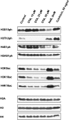
Ochratoxin A (OTA) is one of the most potent rodent renal carcinogens studied to date. Although controversial results regarding OTA genotoxicity have been published, it is now widely accepted that OTA is not a mutagenic, DNA-reactive carcinogen. Instead, increasing evidence from both in vivo and in vitro studies suggests that OTA may promote genomic instability and tumorigenesis through interference with cell division. The aim of the present study was to provide further support for disruption of mitosis as a key event in OTA toxicity and to understand how OTA mediates these effects. Immortalized human kidney epithelial cells (IHKE) were treated with OTA and monitored by differential interference contrast microscopy for 15 h. Image analysis confirmed that OTA at concentrations ≥ 5 μM, which correlate with plasma concentrations in rats under conditions of carcinogenesis, causes sustained mitotic arrest and exit from mitosis without nuclear or cellular division. Mitotic chromosomes were characterized by aberrant condensation and premature sister chromatid separation associated with altered phosphorylation and acetylation of core histones. To test if OTA directly interferes with histone acetyltransferases (HATs) which regulate lysine acetylation of histones and nonhistone proteins, a cell-free HAT activity assay was conducted using total nuclear extracts of IHKE cells. In this assay, OTA significantly blocked HAT activity in a concentration-dependent manner Overall, results from this study provide further support for a mechanism of OTA carcinogenicity involving interference with the mitotic machinery and suggest HATs as a primary cellular target of OTA.
Figures


Similar articles
-
Linking site-specific loss of histone acetylation to repression of gene expression by the mycotoxin ochratoxin A.Arch Toxicol. 2018 Feb;92(2):995-1014. doi: 10.1007/s00204-017-2107-6. Epub 2017 Nov 2. Arch Toxicol. 2018. PMID: 29098329
-
Modulation of key regulators of mitosis linked to chromosomal instability is an early event in ochratoxin A carcinogenicity.Carcinogenesis. 2009 Apr;30(4):711-9. doi: 10.1093/carcin/bgp049. Epub 2009 Feb 23. Carcinogenesis. 2009. PMID: 19237604
-
Ochratoxin A: apoptosis and aberrant exit from mitosis due to perturbation of microtubule dynamics?Toxicol Sci. 2006 Jul;92(1):78-86. doi: 10.1093/toxsci/kfj213. Epub 2006 Apr 26. Toxicol Sci. 2006. PMID: 16641321
-
An update on direct genotoxicity as a molecular mechanism of ochratoxin a carcinogenicity.Chem Res Toxicol. 2012 Feb 20;25(2):252-62. doi: 10.1021/tx200430f. Epub 2011 Nov 16. Chem Res Toxicol. 2012. PMID: 22054007 Review.
-
Ochratoxin A: An overview on toxicity and carcinogenicity in animals and humans.Mol Nutr Food Res. 2007 Jan;51(1):61-99. doi: 10.1002/mnfr.200600137. Mol Nutr Food Res. 2007. PMID: 17195275 Review.
Cited by
-
Human Proximal Tubule Epithelial Cells (HK-2) as a Sensitive In Vitro System for Ochratoxin A Induced Oxidative Stress.Toxins (Basel). 2021 Nov 6;13(11):787. doi: 10.3390/toxins13110787. Toxins (Basel). 2021. PMID: 34822571 Free PMC article.
-
Replication stress: an early key event in ochratoxin a genotoxicity?Arch Toxicol. 2025 Jun;99(6):2577-2594. doi: 10.1007/s00204-025-04004-4. Epub 2025 Mar 10. Arch Toxicol. 2025. PMID: 40063242 Free PMC article.
-
Carcinogens induce loss of the primary cilium in human renal proximal tubular epithelial cells independently of effects on the cell cycle.Am J Physiol Renal Physiol. 2012 Apr 15;302(8):F905-16. doi: 10.1152/ajprenal.00427.2011. Epub 2012 Jan 18. Am J Physiol Renal Physiol. 2012. PMID: 22262483 Free PMC article.
-
Ameliorative Influence of Ajwa Dates on Ochratoxin A-Induced Testis Toxicity.J Microsc Ultrastruct. 2018 Jul-Sep;6(3):134-138. doi: 10.4103/JMAU.JMAU_14_18. J Microsc Ultrastruct. 2018. PMID: 30221139 Free PMC article.
-
Persistence of Epigenomic Effects After Recovery From Repeated Treatment With Two Nephrocarcinogens.Front Genet. 2018 Dec 3;9:558. doi: 10.3389/fgene.2018.00558. eCollection 2018. Front Genet. 2018. PMID: 30559759 Free PMC article.
References
-
- Adler M, Muller K, Rached E, Dekant W, Mally A. Modulation of key regulators of mitosis linked to chromosomal instability is an early event in ochratoxin A carcinogenicity. Carcinogenesis. 2009;30:711–719. - PubMed
-
- Arbillaga L, Vettorazzi A, Gil AG, Joost HM, van Delft JH, Garcia-Jalon JA, Lopez de Cerain A. Gene expression changes induced by ochratoxin A in renal and hepatic tissues of male F344 rat after oral repeated administration. Toxicol. Appl. Pharmacol. 2008;230:197–207. - PubMed
-
- Boorman GA, McDonald MR, Imoto S, Persing R. Renal lesions induced by ochratoxin A exposure in the F344 rat. Toxicol. Pathol. 1992;20:236–245. - PubMed
-
- Carter SL, Eklund AC, Kohane IS, Harris LN, Szallasi Z. A signature of chromosomal instability inferred from gene expression profiles predicts clinical outcome in multiple human cancers. Nat. Genet. 2006;38:1043–1048. - PubMed
Publication types
MeSH terms
Substances
Grants and funding
LinkOut - more resources
Full Text Sources
Molecular Biology Databases

