. 2011 Jun 15;186(12):6762-70.
doi: 10.4049/jimmunol.1003638. Epub 2011 May 6.

Aim2 deficiency in mice suppresses the expression of the inhibitory Fcgamma receptor (FcgammaRIIB) through the induction of the IFN-inducible p202, a lupus susceptibility protein

Affiliations

Aim2 deficiency in mice suppresses the expression of the inhibitory Fcgamma receptor (FcgammaRIIB) through the induction of the IFN-inducible p202, a lupus susceptibility protein

Ravichandran Panchanathan et al. J Immunol. .

Abstract

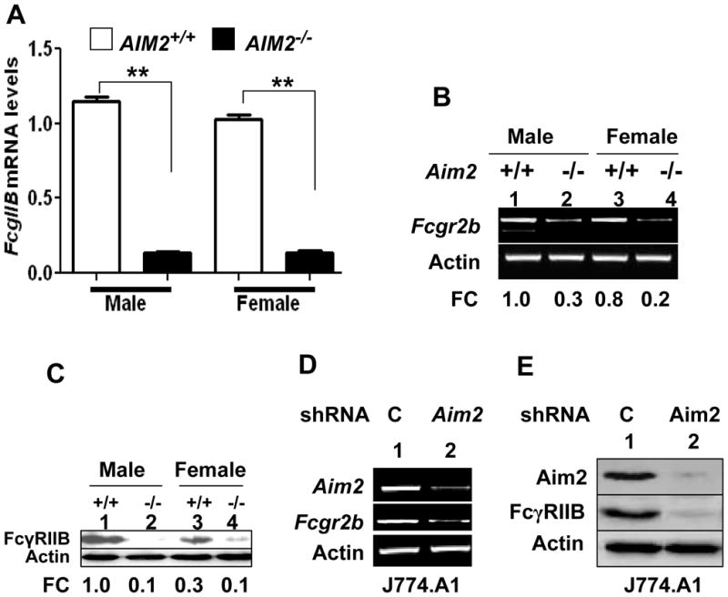
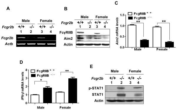
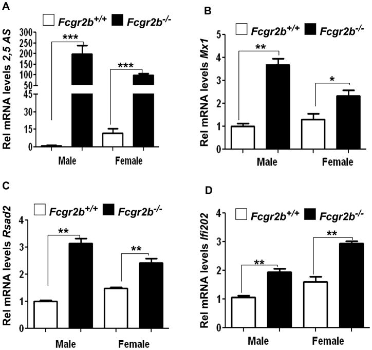
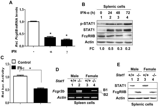
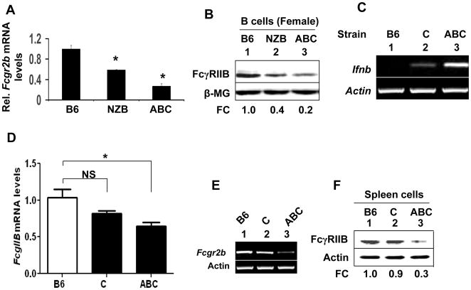
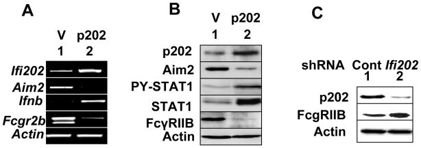
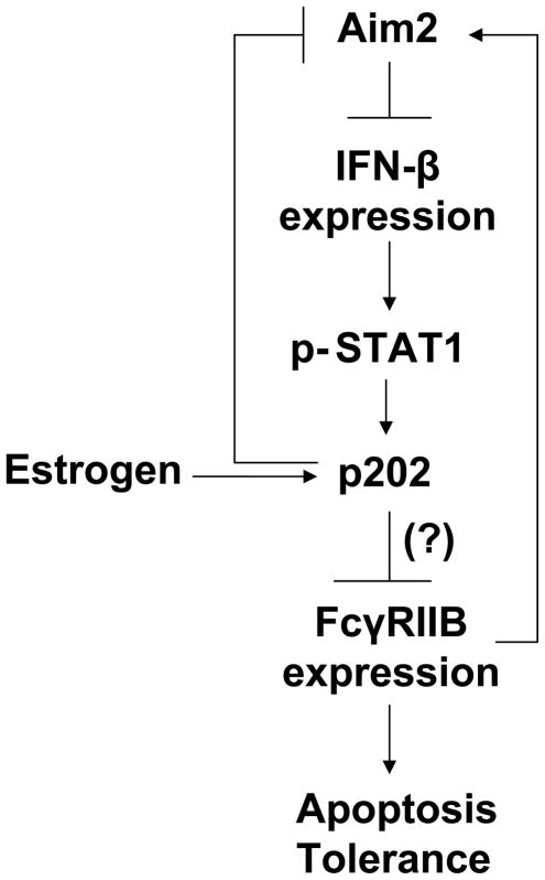
Murine Aim2 and Ifi202 genes (encoding for the Aim2 and p202 proteins) are members of the IFN-inducible Ifi200 gene family. The Aim2 deficiency in mice activates IFN signaling and stimulates the expression of the lupus susceptibility gene, the Ifi202, located within the NZB autoimmunity 2 (Nba2) interval. Given that the deficiency in the expression of the Fcgr2b gene (encoding for the inhibitory FcγRIIB receptor) is associated with increased lupus susceptibility in mice, we investigated whether the Aim2 protein could regulate the expression of Fcgr2b gene. In this article, we report that Aim2 deficiency in mice suppresses the expression of the FcγRIIB receptor. Interestingly, the Fcgr2b-deficient cells expressed increased levels of the IFN-β, activated IFN signaling, and expressed reduced levels of the Aim2 protein. Treatment of splenic cells with IFN-α or -γ reduced levels of the FcγRIIB mRNA and protein and also decreased the activity of the FcγRIIB p(-729/+585) Luc reporter. Moreover, levels of the FcγRIIB receptor were significantly higher in the Stat1-deficient splenic cells than in the wild-type cells. Accordingly, increased expression of IFN-β in lupus-prone B6.Nba2-ABC mice, as compared with non-lupus-prone C57BL/6 (B6) or B6.Nba2-C mice, was associated with reduced expression of the FcγRIIB receptor. Notably, overexpression of the p202 protein in cells decreased the expression of the Aim2 gene, activated the IFN response, and suppressed the expression of the Fcgr2b gene. These observations demonstrate that the expression of Aim2 protein is required to maintain the expression of the Fcgr2b gene and also predict epistatic interactions between the Ifi200 genes and the Fcgr2b gene within the Nba2 interval.


Conflict of interest statement

Disclosures

The authors have no financial conflicts of interest.

Figures

FIGURE 1
FIGURE 1. Aim2-deficiency or knockdown decreases the Fcgr2b expression
(a) Total RNA was extracted from the purified splenic B cells that were isolated from wild-type and age-matched Aim2-deficient male or female mice (age 6–8 weeks). Steady state levels of Fcgr2b mRNA were analyzed by quantitative TaqMan real-time PCR. The ratio of the test gene to β2-microglobulin mRNA was calculated in units (one unit being the ratio of the test gene to β2-microglobulin mRNA). Results are mean values of triplicate experiments and error bars represent standard deviation (** p <0.005). (b) Total RNA as described in panel (a) was also analyzed by semi-quantitative PCR using a pair of primers specific to the indicated genes. (c) Total cell lysates from purified splenic B cells that were prepared from wild-type (lanes 1 and 3) and age-matched Aim2-deficient (lanes 2 and 4) male or female mice (age 6–8 weeks) were analyzed by immunoblotting using antibodies specific to the indicated proteins. The fold change (FC) in the levels of the FcγRIIB receptor protein after normalization with actin protein levels is also indicated. (d) Total RNA was extracted from stably infected with control lentivirus (C) or shAim2 lentivirus (Aim2) J774.A1 cells. The RNA was analyzed by semi-quantitative PCR using a pair of primers specific to the indicated gene. (e) Total cell lysates prepared from J774.A1 cells described in panel (d) were subjected to immunoblotting to detect the indicated proteins.
FIGURE 2
FIGURE 2. Fcgr2b-deficiency induces a type I IFN response
(a) Total RNA was extracted from splenocytes isolated from wild-type (lanes 1 and 3) and age-matched Fcgr2b-deficient (lanes 2 and 4) male or female mice (age ~8 weeks). The RNA was analyzed by semi-quantitative PCR using a pair of primers specific to the indicated genes. (b) Total protein extracts were prepared from splenocytes isolated from wild-type (lanes 1 and 3) and age-matched Fcgr2b-deficient (lanes 2 and 4) male or female mice (age ~8 weeks). Extracts containing approximately equal amounts of proteins were analyzed by immunoblotting for the indicated proteins. (c) The RNA samples described in the panel (a) were subjected to quantitative real-time PCR to detect levels of the Aim2 mRNA. The ratio of the Aim2 mRNA to the β2-microglobulin mRNA was calculated in units (one unit being the ratio of the test gene to β2-microglobulin mRNA). The relative steady-state levels of Aim2 mRNA in male mice are indicated as 1. Results are mean values of triplicate experiments and error bars represent standard deviation (** p <0.01). (d) The RNA samples described in the panel (a) were subjected to quantitative real-time PCR to detect levels of the Ifnb mRNA. The ratio of the Ifnb mRNA to the β2-microglobulin mRNA was calculated in units (one unit being the ratio of the test gene to β2-microglobulin mRNA). The relative steady-state levels of Ifnb mRNA in male mice are indicated as 1. Results are mean values of triplicate experiments and error bars represent standard deviation (* p <0.05; ** p <0.01). (e) Total protein extracts described in panel (b) were analyzed by immunoblotting for the indicated proteins.
FIGURE 3
FIGURE 3. Fcgr2b-deficiency induces the expression of IFN-inducible genes
Total RNA was isolated from total splenocytes isolated from wild-type and age-matched Fcgr2b-deficient male or female mice (age ~8 weeks). Steady state levels of mRNA corresponding to the IFN-inducible 2,5 AS (a), Mx1 (b), Rsad2 (c), and Ifi202 (d) genes were analyzed by quantitative TaqMan real-time PCR. The ratio of the test gene to β2-microglobulin mRNA was calculated in units (one unit being the ratio of the test gene to β2-microglobulin mRNA). Results are mean values of triplicate experiments and error bars represent standard deviation (* p <0.05; ** p <0.005; *** p <0.0005).
FIGURE 4
FIGURE 4. Activation of IFN-signaling decreases FcγRIIB receptor expression
(a) Splenocytes isolated from the B6 female mice (age ~10 weeks) were either left without any treatment or treaded with IFN-α (1,000 u/ml) or IFN-γ (10 ng/ml) for 18 h. Total RNA was extracted after the treatment and analyzed by quantitative real-time PCR for FcγRIIB mRNA levels. The ratio of the FcγRIIB mRNA mRNA to the β2-microglobulin mRNA was calculated in units (one unit being the ratio of the test gene to β2-microglobulin mRNA). The relative steady-state levels of FcγRIIB mRNA in untreated control cells are indicated as 1. Results are mean values of triplicate experiments and error bars represent standard deviation (* p <0.05). (b) Splenocytes isolated from the B6 female mice (age ~10 weeks) were either left without any treatment or treaded with IFN-α (1,000 u/ml) for the indicated time (h). Total cell extracts were subjected to immunoblotting using specific antibodies to the indicated proteins. FC, indicates fold change in the FcγRIIB receptor levels. (c) Cultures of RAW264.7 cells in a 6-well plate were transfected with the FcγRIIB p(−729/+ 585) Luc-reporter plasmid (2.5 μg) along with pRL-TK (0.5 μg) reporter plasmid using FuGENE 6 transfection reagent. 24 h after transfections, cells were either left untreated or treated with IFN-γ. 40–45 h after transfections, cells were processed for the dual luciferase activity. Results are mean values of triplicate experiments and error bars represent standard deviation (* p <0.05). (d) Total RNA was prepared from splenocytes isolated from wild-type (lanes 1 and 3) and age-matched Stat1-deficient (lanes 2 and 4) male or female mice (age ~9 weeks). The RNA was analyzed by semi-quantitative PCR using a pair of primers specific to the indicated genes. (e) Protein extracts containing equal amounts of proteins that were prepared from wild-type (lanes 1 and 3) and age-matched Stat1-deficient (lanes 2 and 4) male or female mice (age ~9 weeks) were subjected to immunoblotting to analyze the levels of the indicated proteins.
FIGURE 5
FIGURE 5. Increased expression of IFN-β in lupus susceptible mice is associated with reduced expression of FcγRIIB receptor
(a) Total RNA isolated from splenocytes prepared from age-matched (~9-weeks) B6, NZB, or B6.Nab2 (ABC) female mice was analyzed by quantitative real-time PCR for FcγRIIB receptor mRNA levels. The ratio of the FcγRIIB mRNA to the β2-microglobulin mRNA was calculated in units (one unit being the ratio of the test gene to β2-microglobulin mRNA). The relative steady-state levels of FcγRIIB mRNA in the B6 female mice are indicated as 1. Results are mean values of triplicate experiments and error bars represent standard deviation (* p <0.05). (b) Total protein extracts from purified B cells (B220+) were subjected to immunoblotting for the indicated protein using the specific antibodies. FC, indicates fold change in the FcγRIIB receptor levels. (c) Total RNA extracted from age-matched (~4-month-old) female B6, B6.Nba2-C (C), or B6.Nba2-ABC (ABC) splenic cells was analyzed by semi-quantitative PCR for Ifnb mRNA levels. (d) Total RNA in the panel (c) was analyzed by quantitative TaqMan real-time PCR using the specific to Fcgr2b. The ratio of the Fcgr2b mRNA levels to β2-microglobulin mRNA levels was calculated in units (one unit being the ratio of the test gene to β2-microglobulin mRNA in splenocytes). The relative levels of Fcgr2b mRNA levels in the B6 females are indicated as 1. Results are mean values of triplicate experiments and error bars represent standard deviation (NS, not significant; * p <0.05). (e) Total RNA in the panel (c) was also analyzed by semi-quantitative PCR for Fcgr2b mRNA levels. (f) Total cell extracts from splenocytes prepared from age-matched (~10-weeks) B6, NZB, or B6.Nab2 (ABC) female mice were analyzed by immunoblotting using antibodies specific to the indicated proteins. FC, indicates fold change in the FcγRIIB receptor levels.
FIGURE 6
FIGURE 6. The expression levels of the p202 protein are inversely correlated with FcγRIIB receptor
(a) Total RNA prepared from RAW264.7 cells either stably transfected with a control vector (pCMV; indicated as V) or a plasmid (pCMV-202; indicated as p202) that allowed the expression of the Ifi202 gene was analyzed by semi-quantitative PCR using a pair of primers specific to the indicated genes. (b) Total cell extracts from RAW264.7 cell clones described in the panel (a) were analyzed by immunoblotting using antibodies specific to the indicated proteins. (c) Total cell extracts from J774.A1 cells infected with control lentivirus (lane 1) or shIfi202 lentivirus (lane 2) were subjected to immunoblotting for the indicated proteins.
FIGURE 7
FIGURE 7. Proposed regulatory role of the Aim2 protein in the maintenance of the FcγRIIB receptor expression through down-regulation of the p202 protein expression

Similar articles

Cited by

References

    1. Graham RR, Hom G, Ortmann W, Behrens TW. Review of recent genome-wide association scans in lupus. J Intern Med. 2009;265:680–688. - PubMed
    1. Moser KL, Kelly JA, Lessard CJ, Harley JB. Recent insights into the genetic basis of systemic lupus erythematosus. Genes Immun. 2009;10:373–379. - PMC - PubMed
    1. Kono DH, Theofilopoulos AN. Genetics of SLE in mice. Springer Semin Immunopathol. 2006;28:83–96. - PubMed
    1. Morel L. Genetics of SLE: evidence from mouse models. Nat Rev Rheumatol. 2010;6:348–357. - PubMed
    1. Cohen-Solal JF, Jeganathan V, Grimaldi CM, Peeva E, Diamond B. Sex hormones and SLE: influencing the fate of autoreactive B cells. Curr Top Microbiol Immunol. 2006;305:67–88. - PubMed

Publication types

MeSH terms