. 2011 Jul 11;52(8):5144-52.
doi: 10.1167/iovs.10-7047.

Preventive effects of ethyl pyruvate on endotoxin-induced uveitis in rats

Affiliations

Preventive effects of ethyl pyruvate on endotoxin-induced uveitis in rats

Nilesh M Kalariya et al. Invest Ophthalmol Vis Sci. .

Abstract

Purpose: Recent studies indicate that ethyl pyruvate (EP) exerts anti-inflammatory properties; however, the effect of EP on ocular inflammation is not known. The efficacy of EP in endotoxin-induced uveitis (EIU) in rats was investigated.

Methods: EIU in Lewis rats was developed by the subcutaneous injection of lipopolysaccharide (LPS; 150 μg). EP (30 mg/kg body weight) or its carrier was injected intraperitoneally 1 hour before or 2 hours after lipopolysaccharide injection. Animals were killed after 3 and 24 hours followed by enucleation of eyes and collection of the aqueous humor (AqH). The number of infiltrating cells and levels of proteins in the AqH were determined. The rat cytokine/chemokine multiplex method was used to determine level of cytokines and chemokines in the AqH. TNF-α and phospho-nuclear factor kappa B (NF-κB) expression in ocular tissues were determined immunohistochemically. Human primary nonpigmented ciliary epithelial cells (HNPECs) were used to determine the in vitro efficacy of EP on lipopolysaccharide-induced inflammatory response.

Results: Compared to controls, AqH from the EIU rat eyes had a significantly higher number of infiltrating cells, total protein, and inflammatory cytokines/chemokines, and the treatment of EP prevented EIU-induced increases. In addition, EP also prevented the expression of TNF-α and activation of NF-κB in the ciliary bodies and retina of the eye. Moreover, in HNPECs, EP inhibited lipopolysaccharide-induced activation of NF-κB and expression of Cox-2, inducible nitric oxide synthase, and TNF-α.

Conclusions: Our results indicate that EP prevents ocular inflammation in EIU, suggesting that the supplementation of EP could be a novel approach for the treatment of ocular inflammation, specifically uveitis.


Figures

Figure 1.
Figure 1.
Structure of ethyl pyruvate.
Figure 2.
Figure 2.
EP prevents LPS-induced inflammatory cell infiltration and increase in protein concentration in rat AqH. (A–C) LPS was injected in rats pretreated (1 hour before LPS injection) without or with EP (30 mg/kg body weight) to induce uveitis. (A) Serial sections of paraformaldehyde-fixed rat eyes enucleated 24 hours after EIU-induction were stained with hematoxylin–eosin and were observed under a light microscope to examine the infiltration of inflammatory cells in the anterior chamber. (B) The infiltrated inflammatory cells and (C) total protein concentration in the rat AqH at 24 hours. (D and E) Uveitis was induced in rats by injecting with LPS followed by treatment (2 hours after LPS injection) without or with EP (30 mg/kg body weight). (D) The infiltrated inflammatory cells and (E) total protein concentration in the rat AqH at 24 hours. (F and G) LPS was injected in rats pretreated (1 hour before LPS) without or with EP (0, 15, 30, and 60 mg/kg body weight) to examine the dose-dependent effects of EP on EIU. (F) The infiltrated inflammatory cells and (G) total protein concentration in the rat AqH at 24 hours. Trypan blue exclusion cell counting method was used to determine number of infiltrating cells, and the Bradford method was used to measure total protein concentration. Results are expressed as the mean ± SD (n = 6). *P < 0.001 vs. the control group; **P < 0.01 vs. the EIU group. Magnification: (A), ×200.
Figure 3.
Figure 3.
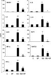
EP Inhibits LPS-induced increase in cytokines and chemokines in EIU eye AqH. Inflammatory cytokine and chemokine levels in the AqH collected 24 hours after LPS injection were measured by using the MILLIPLEX MAG rat cytokine/chemokine magnetic bead panel as described in the methods. Results are expressed as the mean ± SE (n = 3; AqH was pooled from 2 rats for each data point); *P < 0.001 or **P < 0.01 vs. the control group; *P < 0.001 or **P < 0.01 vs. EIU group. The results were analyzed and expressed in picograms per milliliter.
Figure 4.
Figure 4.
EP suppresses the expression of TNF-α in ocular tissues of EIU rats. Serial sections of paraformaldehyde-fixed rat eyes enucleated 24 hours after EIU were immunostained with DAPI and antibodies against TNF-α. The DAPI and antibody staining intensity was observed by fluorescent microscope. Representative images are shown (n = 4). C, Control; EP, ethyl pyruvate; EIU, endotoxin-induced uveitis; EIU + EP, endotoxin-induced uveitis + ethyl pyruvate; IMNC, isotype-matched negative control. Magnification: ×400.
Figure 5.
Figure 5.
EP suppresses the activation of NF-κB in ocular tissues of LPS-induced EIU. Serial sections of paraformaldehyde-fixed rat eyes enucleated 3 hours after EIU induction were immunostained with DAPI and antibodies against phospho-p65. The DAPI and antibody staining intensity was observed by fluorescent microscope. Representative images are shown (n = 4). C, Control; EP, ethyl pyruvate; EIU, endotoxin-induced uveitis; EIU + EP, endotoxin-induced uveitis + ethyl pyruvate; IMNC, isotype-matched negative control. Magnification: ×400.
Figure 6.
Figure 6.
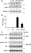
EP prevents inflammatory response in HNPECs stimulated with LPS. (A) Growth-arrested HNPECs pretreated without or with EP (40 μM) were incubated with 1 μg/mL of LPS for 24 hours. The expression of iNOS and Cox-2 from cell lysates was determined by Western blot analysis using specific antibodies. (B) The levels of TNF-α in the culture media was determined with ELISA kit. Data are expressed as the mean ± SD (n = 6). *P < 0.001 vs. the control group; **P < 0.01 vs. the LPS group. (C) Growth-arrested HNPECs pretreated with or without EP (40 μM) were incubated with 1 μg/mL of LPS for 0 to 90 minutes. The expression levels of phospho- and total-p65 were determined by Western blot analysis using specific antibodies.
Figure 7.
Figure 7.
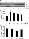
EP prevents LPS-induced nuclear translocation of p65, ROS generation and HNPECs death. (A) Growth-arrested HNPECs pretreated with or without EP (40 μM) were incubated with 1 μg/mL of LPS for 0 to 60 minutes The translocation of p65 from cytoplasm to nucleus was determined by Western blot analysis using specific antibodies. (B) Growth-arrested HNPECs pretreated with or without EP (10, 20, and 40 μM) were incubated with 5 μg/mL of LPS for 1 hour. The ROS measurement was performed using H2DCFDA dye. (C) Growth-arrested HNPECs pretreated with or without EP (40 μM) were incubated with 5 μg/mL of LPS for 24 hours. The cell viability was determined by MTT assay. Data are expressed as the mean ± SD (n = 4). *P < 0.001 vs. the control group; **P < 0.01 vs. the LPS group. EP, Ethyl pyruvate.

Similar articles

Cited by

References

    1. Holleman MAF. Notice sur l'action de l'eau oxygénée sur les acétoniques et sur le dicétones. Recl Trav Chim Pays-bas Belg. 1904;23:169–171
    1. Ervens B, Gligorovski S, Herrmann H. Temperature dependent rate constants for hydroxyl radical reactions with organic compounds in aqueous solutions. Phys Chem Chem Phys. 2003;5:1811–1824
    1. Varma SD, Hegde KR. Lens thiol depletion by peroxynitrite. Protective effect of pyruvate. Mol Cell Biochem. 2007;298:199–204 - PubMed
    1. Kao KK, Fink MP. The biochemical basis for the anti-inflammatory and cytoprotective actions of ethyl pyruvate and related compounds. Biochem Pharmacol. 2010;80:151–159 - PubMed
    1. Yang R, Gallo DJ, Baust JJ, et al. Ethyl pyruvate modulates inflammatory gene expression in mice subjected to hemorrhagic shock. Am J Physiol Gastrointest Liver Physiol. 2002;283:G212–G222 - PubMed

Publication types

MeSH terms