Comparative Study
. 2011 Jun;8(6):499-508.
doi: 10.1038/nmeth.1605. Epub 2011 May 8.

Fast, three-dimensional super-resolution imaging of live cells

Affiliations
Comparative Study

Fast, three-dimensional super-resolution imaging of live cells

Sara A Jones et al. Nat Methods. 2011 Jun.

Abstract

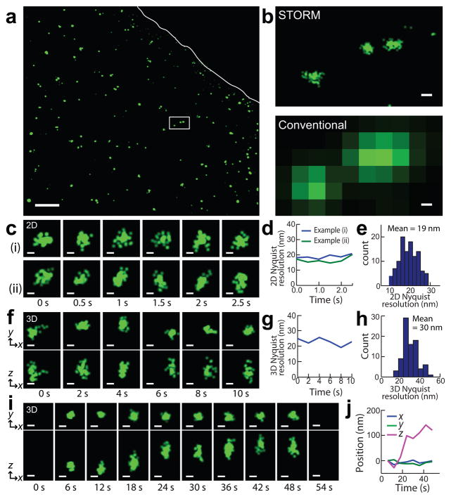
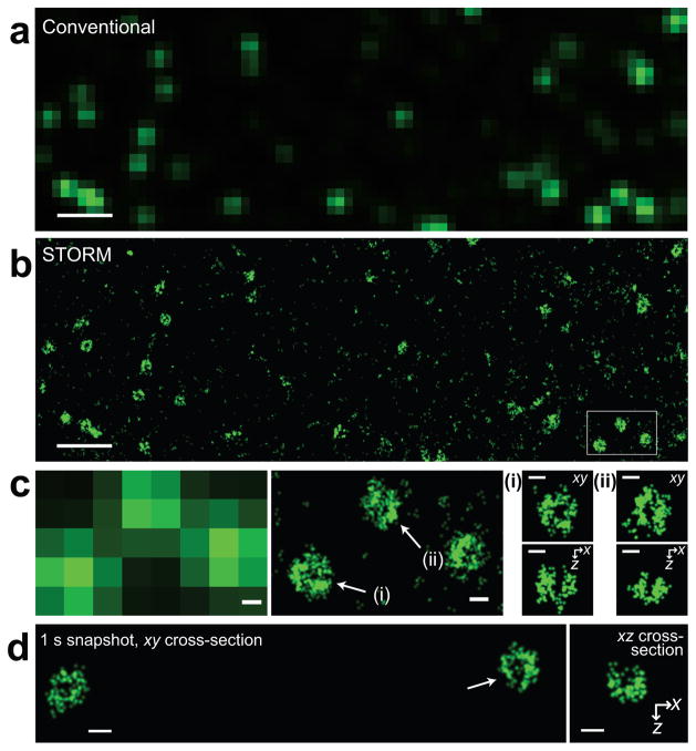
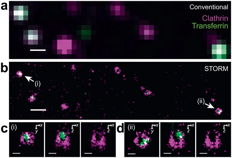
We report super-resolution fluorescence imaging of live cells with high spatiotemporal resolution using stochastic optical reconstruction microscopy (STORM). By labeling proteins either directly or via SNAP tags with photoswitchable dyes, we obtained two-dimensional (2D) and 3D super-resolution images of living cells, using clathrin-coated pits and the transferrin cargo as model systems. Bright, fast-switching probes enabled us to achieve 2D imaging at spatial resolutions of ∼25 nm and temporal resolutions as fast as 0.5 s. We also demonstrated live-cell 3D super-resolution imaging. We obtained 3D spatial resolution of ∼30 nm in the lateral direction and ∼50 nm in the axial direction at time resolutions as fast as 1-2 s with several independent snapshots. Using photoswitchable dyes with distinct emission wavelengths, we also demonstrated two-color 3D super-resolution imaging in live cells. These imaging capabilities open a new window for characterizing cellular structures in living cells at the ultrastructural level.


Conflict of interest statement

Competing interests statement.

The authors declare that they have no competing financial interests.

Figures

Figure 1
Figure 1. STORM images of transferrin in live cells
(a) A wide-field 2D STORM image. Analysis was limited to inside the cell boundary, demarcated by the white line, due to unreliable STORM localizations outside the cell caused by fluorescence background from diffusing transferring molecules. (b) The zoomed-in comparison between 2D STORM (upper panel) and conventional (lower panel) images of the boxed region in (a). (c) Examples of two transferrin clusters, each showing six consecutive 2D STORM snapshots (0.5 sec per snapshot). (d) The 2D Nyquist resolution for the examples shown in (c). (e) A histogram of Nyquist resolutions constructed from many individual transferrin clusters at 0.5-sec time resolution. (f) Consecutive 3D STORM snapshots of a transferrin cluster (2 sec per snapshot). Upper row: xy projections. Lower row: xz projections. (g) The 3D Nyquist resolution derived from the snapshots in (f). (h) A histogram of 3D Nyquist resolutions, constructed from many clusters at 2-sec time resolution. (i) Images of a transferrin cluster at 6 sec/snapshot at 34°C showing its time evolution, starting from formation (6 sec) to disappearance (54 sec) upon internalization. Upper row: xy projections. Lower row: xz projections. (j) The x, y and z coordinates of the centroid position of the transferrin cluster in (i). Scale bars, 2 μm (a), 100 nm (b), 50 nm (c,f,i). In all figures, the z axis points away from the glass substrate.
Figure 2
Figure 2. 3D STORM images of clathrin-coated pits (CCPs) labeled with photoswitchable cyanine dyes via a SNAP tag in live cells
(a) Conventional wide-field image of CCPs in a live cell. (b) 3D STORM image of the same area taken in 30 seconds. Here, only the xy projection is shown. (c) Zoomed-in conventional (left panel) and 3D STORM (center panel) images of the boxed region in (b). The 3D STORM image is again presented as the xy projection. Right panels: Cross-sections of the CCPs indicated in the center panel. Upper row: an xy cross-section near the plasma membrane. Lower row: an xz cross-section cutting through the middle of the pit. (d) 3D STORM image of CCPs taken in 1 second. Left panel: xy cross-section of two CCPs. Right panel: xz cross-section of the CCP indicated by the arrow. Scale bars, 1 μm (a,b), 100 nm (c,d).
Figure 3
Figure 3. Two-color 3D STORM images of CCPs and transferrin in live cells
CCPs are labeled with Alexa647 via a SNAP tag (magenta) and transferrin is directly labeled with Alexa568 (green). (a) Conventional image of CCPs and transferrin in a live cell. (b) 3D STORM image of the same area taken in 30 seconds. The xy projection of the 3D image is displayed. (c,d) Zoomed-in STORM images of the CCPs indicated in (b). Left panel: the xy cross-section near the plasma membrane. Center panel: xz cross-section cutting through the middle of the invaginating pit. Right panel: corresponding xz cross-section of the clathrin channel only. Scale bars, 500 nm (a,b), 100 nm (c,d).
Figure 4
Figure 4. Comparison of CCPs labeled with various probes
Composite CCPs constructed by aligning the 3D center-of-mass of CCPs in an entire field of view for each probe. A portion of the field of view is displayed in Fig. 2b for Alexa647 and in Supplementary Fig. 8 for tdEos, mEos2, Atto655, TMR and Oregon Green. The xy cross-sections through the 3D center of the composite CCPs are displayed in the upper panels. The localization density profiles along the x axis within a 75-nm wide stripe at the center of the pits are shown in the lower panels. Scale bar, 100 nm.

References

    1. Hell SW. Far-field optical nanoscopy. Science. 2007;316:1153–1158. - PubMed
    1. Huang B, Babcock H, Zhuang X. Breaking the diffraction barrier: super-resolution imaging of cells. Cell. 2010;143:1047–1058. - PMC - PubMed
    1. Klar TA, Hell SW. Subdiffraction resolution in far-field fluorescence microscopy. Opt Lett. 1999;24:954–956. - PubMed
    1. Heintzmann R, Jovin TM, Cremer C. Saturated patterned excitation microscopy -a concept for optical resolution improvement. J Opt Soc Am A. 2002;19:1599–1609. - PubMed
    1. Gustafsson MGL. Nonlinear structured-illumination microscopy: Wide-field fluorescence imaging with theoretically unlimited resolution. Proc Natl Acad Sci U S A. 2005;102:13081–13086. - PMC - PubMed

Publication types

MeSH terms