. 2011 Jun;18(6):650-7.
doi: 10.1038/nsmb.2032. Epub 2011 May 8.

Structure of C3PO and mechanism of human RISC activation

Affiliations

Structure of C3PO and mechanism of human RISC activation

Xuecheng Ye et al. Nat Struct Mol Biol. 2011 Jun.

Abstract

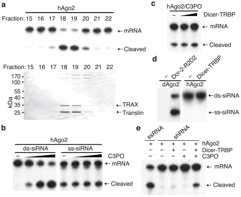
Assembly of the RNA-induced silencing complex (RISC) consists of loading duplex (guide-passenger) siRNA onto Argonaute (Ago2) and removing the passenger strand. Ago2 contributes critically to RISC activation by nicking the passenger strand. Here we reconstituted duplex siRNA-initiated RISC activity using recombinant human Ago2 (hAgo2) and C3PO, indicating that C3PO has a critical role in hAgo2-RISC activation. Consistently, genetic depletion of C3PO compromised RNA silencing in mammalian cells. We determined the crystal structure of hC3PO, which reveals an asymmetric octamer barrel consisting of six translin and two TRAX subunits. This asymmetric assembly is critical for the function of C3PO as an endonuclease that cleaves RNA at the interior surface. The current work supports a Dicer-independent mechanism for human RISC activation, in which Ago2 directly binds duplex siRNA and nicks the passenger strand, and then C3PO activates RISC by degrading the Ago2-nicked passenger strand.


Figures

Figure 1
Figure 1. Reconstitution of human RISC activity
(a) The duplex siRNA-initiated RISC assays (top) and colloidal stained SDS-PAGE gel (bottom) showing the purified RISC activation activity and correlated proteins. (b) Comparison of C3PO’s RISC activation activities using duplex (ds)-siRNA (left 4 lanes) and single-stranded (ss)-siRNA (right 4 lanes) as triggers. While Ago2 concentration was ~0.1 μM, the titration of C3PO was ~0.01, 0.03, and 0.09 μM in this experiment. (c) The duplex siRNA-initiated RISC assays comparing the RISC activities using recombinant hAgo2 and hC3PO in the absence or presence of Dicer-TRBP complex. The relative concentration of Ago2:C3PO:Dicer-TRBP is approximately 1:1:1. (d) Native PAGE gel showing the siRNAs associated with recombinant dAgo2 or hAgo2 in the absence or presence of, respectively, the Dcr-2-R2D2 or Dicer-TRBP complex. (e) Reconstitution of shRNA-initiated RISC activity with various combinations of recombinant Dicer-TRBP complex, hAgo2, and hC3PO.
Figure 2
Figure 2. C3PO is required for efficient RNAi in mammalian cells
(a) Western blots comparing the levels of endogenous Ago2, Translin, or Actin proteins in wild type, trsn−/−, and trsn−/− (Flag-Translin) MEF cells. (b) Comparison of the duplex siRNA-initiated RISC activities of the cell lysates prepared from wild-type, trsn−/−, and trsn−/− (Flag-Translin) MEFs. Protein concentrations were equalized prior to the assays. (c) The deficiency of RISC activity in trsn−/− cell extract could be efficiently rescued by addition of recombinant C3PO. (d) A schematic showing the comparison of the in vivo siRNA efficiencies between wild-type, trsn−/−, and trsn−/− (Flag-Translin) MEF cells using different concentration of siRNA. Error bars indicate standard deviations by multiple experiments.
Figure 3
Figure 3. Crystal structure of C3PO
(a) Ribbon representation of C3PO complex, in which the two TRAX subunits are colored in yellow and orange, and the six Translin subunits are colored in cyan (top) or light cyan (bottom). (b) An orthogonal view of (a) from the top of C3PO complex. (c) Ribbon representation of human TRAX monomer color ramped from N-terminus (blue) to C-terminus (red). The seven helices in the structure are labeled (α1–α7). (d) Ribbon representation of the TRAX-Translin heterodimer. Two close-up views illustrate in detail the conserved hydrophobic interactions between the α1 helix of TRAX (or Translin) and the α5 and α6 helices of Translin (or TRAX). The bottom close-up view was rotated for 180 degree to compare with the top one. (e) Packing of two TRAX-Translin heterodimer in C3PO complex. The two TRAX subunits contact each other mainly through the α1 helix. (f) A close-up view of the salt bridge network (dotted lines) between the α1 helices of two adjacent TRAX subunits (left). A Translin-Translin interface is shown in comparison (right), illustrating the varied interactions between the two types of interfaces. The distances between selected residues on Translin are also indicated.
Figure 4
Figure 4. Asymmetric octameric assembly of C3PO
(a) (Left) A schematic model of C3PO structure illustrating the superhelical shift among the top four (TRAX and three Translin) subunits. (Right) A close-up view of specific interactions between TRAX and Translin subunits near the C-terminus. Arg263 of TRAX (1) forms salt bridges throughout with Asp211 and Glu207 of Translin (4), and Glu266 of TRAX (1) interacts with Arg215 of Translin (3). The black arrow indicates the vertical shift between TRAX (1) and Translin (4). The pink arrow indicates the right-handed spiral movement from TRAX (1) to Translin (4). (b) A profiles overlay of Supdex 200 gel filtration chromatography of recombinant wild type (WT), R263E, E207A, D211A, and E207A/D211A mutant C3PO complexes. (c) The native gel shift assays comparing the ability of recombinant wild type and mutant C3PO complexes to bind 5′-radiolabelled ss-siRNA. (d) Comparison of the nuclease activities of recombinant wild type or mutant C3PO complexes.
Figure 5
Figure 5. Catalytic center of C3PO
(a) The anomalous difference electron density map unambiguously reveals the locations of a single Mn2+ ion as well as two SO42− and two PO43− ions (distinguished by the presence of small anomalous difference peaks at the sulfur positions). The catalytic residues (E126, E129, D193 and E197 of TRAX) cluster around the Mn2+, which coordinates E129 and E197 of TRAX and one SO42− ion. (b) The native gel shift assays comparing the ss-siRNA binding abilities between wild type and four catalytic mutants (E126A, E129A, D193A, E197A) of C3PO. (c) Comparison of the nuclease activities between wild type C3PO and catalytic mutants. (d) A model of ssRNA bound to the active site of C3PO. In this model, the bases of ssRNA point outward, whereas the backbone phosphates directly contact several positive-charged residues, such as R192 of Translin and K68, R200 of TRAX. The red arrow indicates the phosphoester bond that is to be cleaved to generate 5′-phosphate and 3′-hydroxyl products. (e) The native gel shift assays showing the comparison of the ss-siRNA binding abilities between wild type and three mutants (K68A, R192A, R200A) of C3PO. (f) Comparison of the nuclease activities between wild type C3PO and RNA-binding mutants.
Figure 6
Figure 6. C3PO cleaves ssRNA at the interior surface
(a) The Superdex 200 gel filtration profiles of ss-siRNA, recombinant C3PO, a C3PO-ss-siRNA mixture in the presence of 5 mM EDTA (to block catalysis, but not RNA binding). (b) A silver stained Urea-PAGE showing the presence of ss-siRNA in the fractions of Superdex 200 gel filtration chromatography from (a). (c) A cylindrical cartoon representation of human C3PO hetero-octamer. The two TRAX subunits (Position 1) are colored yellow and orange respectively; each Translin subunit is colored differently according to its specific position in the complex. The positions of the four subunits at the top of the barrel are labeled on their C-terminal α7 helices, showing the relative vertical shift between each subunit. The ssRNA binding and catalytic residues of C3PO are located at the inside of the barrel highlighted by magenta. (d) The electrostatic potential mapped on the inside (upper) and outside (lower) molecular surface of C3PO. Negatively potentials are shown in red and positively potentials are in blue. The modeled ssRNA strands are shown in green cartoons. Arrows indicate the positions of the two catalytic sites.
Figure 7
Figure 7. C3PO activates human RISC by degrading Ago2-nicked passenger strand
(a) The duplex siRNA-initiated RISC assays comparing the RISC activation activities between recombinant wild type and four catalytic mutants of C3PO. (b) Comparison of the RISC activation activities between recombinant wild type and three RNA-binding mutants of C3PO. (c) The nuclease assays showing the cleavage of duplex siRNA and various nicked duplex siRNAs containing 9-nt and/or 12-nt passenger strand fragments by recombinant C3PO℗ indicates the 5′-radiolabelling. (d) Comparison of the RISC activities of recombinant hAgo2 with or without C3PO using a perfect duplex siRNA, a nicked duplex siRNA, and a nicked duplex siRNA carrying 2′-O-methylated passenger fragments as triggers. indicates 2′-O-methylated RNA.

References

    1. Siomi H, Siomi MC. On the road to reading the RNA-interference code. Nature. 2009;457:396–404. - PubMed
    1. Liu Q, Paroo Z. Biochemical principles of small RNA pathways. Annu Rev Biochem. 2010;79:295–319. - PubMed
    1. Carthew RW, Sontheimer EJ. Origins and Mechanisms of miRNAs and siRNAs. Cell. 2009;136:642–55. - PMC - PubMed
    1. Kawamata T, Tomari Y. Trends Biochem Sci. Making RISC. - PubMed
    1. Liu J, et al. Argonaute2 is the catalytic engine of mammalian RNAi. Science. 2004;305:1437–41. - PubMed

Publication types

MeSH terms