. 2011 Jun;18(6):665-72.
doi: 10.1038/nsmb.2049. Epub 2011 May 8.

Nemo kinase phosphorylates β-catenin to promote ommatidial rotation and connects core PCP factors to E-cadherin-β-catenin

Affiliations

Nemo kinase phosphorylates β-catenin to promote ommatidial rotation and connects core PCP factors to E-cadherin-β-catenin

Ivana Mirkovic et al. Nat Struct Mol Biol. 2011 Jun.

Abstract

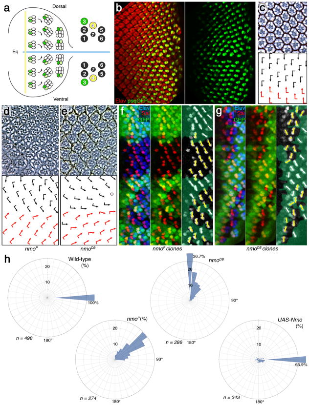
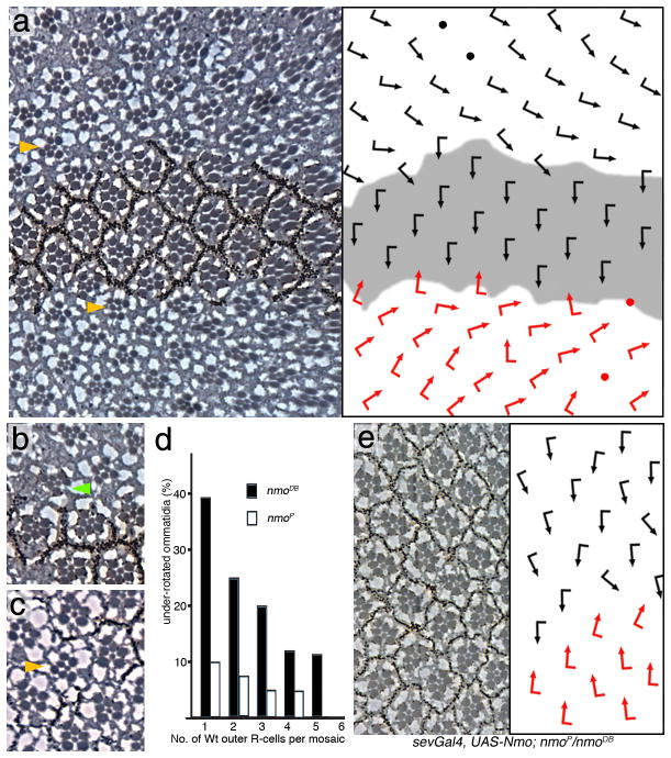
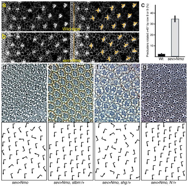
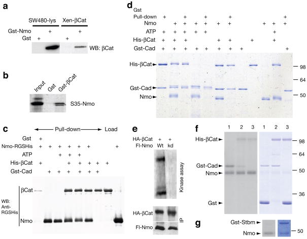
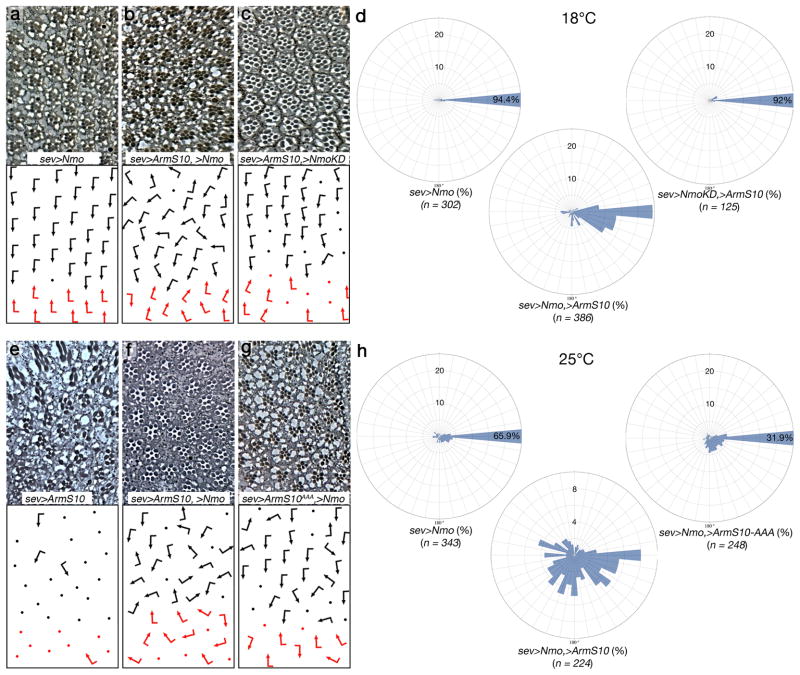
Frizzled planar cell polarity (PCP) signaling regulates cell motility in several tissues, including ommatidial rotation in Drosophila melanogaster. The Nemo kinase (Nlk in vertebrates) has also been linked to cell-motility regulation and ommatidial rotation but its mechanistic role(s) during rotation remain obscure. We show that nemo functions throughout the entire rotation movement, increasing the rotation rate. Genetic and molecular studies indicate that Nemo binds both the core PCP factor complex of Strabismus-Prickle, as well as the E-cadherin-β-catenin (E-cadherin-Armadillo in Drosophila) complex. These two complexes colocalize and, like Nemo, also promote rotation. Strabismus (also called Vang) binds and stabilizes Nemo asymmetrically within the ommatidial precluster; Nemo and β-catenin then act synergistically to promote rotation, which is mediated in vivo by Nemo's phosphorylation of β-catenin. Our data suggest that Nemo serves as a conserved molecular link between core PCP factors and E-cadherin-β-catenin complexes, promoting cell motility.


Figures

Figure 1
Figure 1. nmo is required throughout ommatidial rotation
(a) Schematic presentation of ommatidial rotation in 3rd instar larval eye disc, posterior to the morphogenetic furrow (MF, vertical yellow line; equator, horizontal blue line). Cells acquiring R3 fate are labeled in green. Left panel shows organization of individual photoreceptors within an ommatidium in adult eye. (b) 3rd instar eye disc stained with antibody against a pan-neuronal marker, Elav (red) and psq>GFP reporter (green; strong in R3 and weaker in R4; single channel in right panel). (c) Tangential section of adult wild-type eye with ommatidia arranged around the equator (having completed the 90° rotation). Bottom panel: schematic with dorsal and ventral chiral forms indicated by black and red arrows, respectively. (de) Eye sections of the hypomorphic allele nmoP (d) and the null allele nmoDB (e). Arrows indicate degree of rotation (see quantifications of rotation angle distribution in panel h). (fg) 3rd instar eye discs (just posterior to MF) stained with anti-Sal (red; marking R3/R4 precursors in rows 2–5), anti-Elav (blue; all R-cell precursors) and anti-GFP (green; mutant tissue marked by GFP absence). Mutant clones of nmoP (f) and nmoDB (g) are shown. Right panels: semi-schematic versions of middle panels of f and g, white bars indicating orientation of Wt clusters and yellow bars indicate orientation of mutant clusters. (h) Rose diagrams displaying the angle distribution of ommatidia (in interval of 10°) of the genotypes indicated. The radial axis displays % (up to 25%), percentages above 25% are written next to sector-bar.
Figure 2
Figure 2. nmo is required in a cluster autonomous manner for rotation
Panels ac, and e show tangential sections of adult eyes, (d) shows a quantification; dorsal is up and anterior left. The respective schematic presentations of ommatidial orientation (arrows as in Fig. 1) are shown as right panels in a and e. (ac) Mutant clones of nmoDB, marked by absence of pigment (Wt area in schematic in a is shaded gray), In a: examples of non-rotated ommatidia adjacent to wt tissue are highlighted by yellow arrowheads. (b) Green arrowhead marks an under-rotated mosaic ommatidium. (c) A mutant ommatidium (yellow arrowhead) surrounded by wild-type clusters. (d) Quantification of rotation in mosaic ommatidia: (nmoDB is shown with black bars and nmoP in white bars). (e) sev>Nmo; nmoP/nmoDB. Expression of Nmo protein in a subset of R-cells in flies homozygous mutant for nmo.
Figure 3
Figure 3. Increased Nmo levels cause an increased rotation rate
(ab) Confocal microscopy images of 3rd instar eye imaginal discs, anterior is left and dorsal is up; (a) wild-type and (b) sevGal4, UAS-Nmo (sev>Nmo). Discs are stained with anti-Arm, labeling all cell membranes. Arm is enriched on membranes within the forming cluster. Yellow bars delineate rotation angles of preclusters. (c) Quantification of rotation angles in rows up to row 9 in % +/− s.d. . (dg) Adult eyes of sev>Nemo genotypes as indicated, dorsal area of eye is shown, anterior is left (see Table 1 for quantification). (d) sev>Nmo (in w1118 background). (e) sev>Nemo, stbm/+. (f) sev>Nemo, shg/+. (g) sev>Nemo, N/+. Other genotypes: see Table 1.
Figure 4
Figure 4. Nemo interacts physically and genetically with Stbm and Pk
(a) Gst-pulldown assays using Gst-Nmo full length and in vitro translated StbmC, the part common to all Pk isoforms (PkC), Dsh, Dgo and PkM (b) Specificity assays for the Gst-Nmo/StbmC interaction, using Gst-PkC: ref. The “input” lane is 10%. (cf) Tangential eye sections of stbm6 (panel c), stbm6; nmoP (d), pksple1, nmoP (e) and pksple1 (f). Schematic presentation of cluster orientation is shown below each micrograph; arrows are is in Fig. 1. (gh) Precluster rotation defects in pksple1, nmoP double mutant discs (CnoGFP is used to mark all cells at the adherens junctions). Orientation of clusters is highlighted by orange bars; orange arrows highlight one cluster each in g and h that have not yet initiated rotation. (ik) NmoGFP is localized to the membrane at the R4 side of the R3/R4 border. (i) Area of eye imaginal disc posterior to furrow (anterior is left, dorsal up) showing NmoGFP localization (green) and membrane staining (anti-Arm; magenta). Boxes indicate areas shown at high magnification in j and k, respectively. (jk) High magnifications of preclusters that initiated rotation (j) and one that is approximately at a 45° (k). R4 side of the R3/R4 border in both clusters is highlighted by yellow arrowheads, also in panel i; the 5 R-cells of the precluster are labeled by their numbers; bottom panels: monochromes showing NmoGFP only with a semi-schematic overlay of cell outlines (orange). R4-side of membrane is indicated by black arrowheads. (lm) NmoGFP localization and stability depends on the presence of Stbm. (l) NmoGFP localization in wild-type background. R3 and R4 precursors are labeled with 3 and 4, respectively, within each cluster. (m) In GMR>NmoGFP; >stbmRNAi the respective mutant R3/R4 precursors are marked with stars. Anterior is left and dorsal up in both panels.
Figure 5
Figure 5. Nemo binds and phosphorylates β-catenin and Cadherin
(a) Extracts from SW40 cell lysates incubated with Gst-Nemo. β-catenin is detected with antiβcat (left panel). Recombinant Xenopus β-cat incubated with Gst-Nmo (right panel). (b) Gst–βcat (Arm) incubated with in vitro translated Nmo. (c) Western blot with RGS-His antibody detection of the respective Gst-Cad pulldowns as indicated. (d) Coomassie stained gel of Gst-Cad (C-cadherin) pull-down assays and kinase reactions with Nmo-His and His-β-cat (in the presence or absence of ATP). (eg) 32P kinase reactions with Nmo. (e) Nmo purified from cell lysates: full length Nemo (Nmo-FL) wild-type protein (lane 1; Wt) or kinase inactive (lane 2; kd) and β-cat (Arm; upper band). (fg) In vitro kinase reactions with bacculovirus expressed, purified Nmo. In f, lanes 1: Nmo, Gst-Cad, and Gst; lanes 2: Nmo, Gst-Cad, and His-β–catenin: and lanes 3: Nmo, His-β–catenin, and Gst. In g, Nmo and Gst-Stbm. Left panels show radioactive exposures, revealing kinase events of respective Coomassie stained proteins shown on right in panels (f) and (g).
Figure 6
Figure 6. The activity of Arm/β-catenin is modulated by Nmo in vivo
(ac) and (eg) Tangential eye sections of genotypes indicated, anterior is left and dorsal up. Ommatidial orientation is presented schematically in lower panels (arrows are as in Fig. 1, dots represent ommatidia that cannot be scored for orientation). (ac) Expression of indicated transgenes at 18°C under sevGal4 control; (a) UAS-Nmo, (b) UAS-Nmo, UAS-ArmS10 (a stabilized form of Arm/β-cat) (c) UAS-ArmS10, UAS-NmoKD (a kinase inactive isoform). (d) Quantification of rotational enhancements: rose diagrams in 10° intervals of the genotypes indicated expressed at 18°C (corresponding to the genotypes shown in panels ac). The radial axis displays % (up to 25%), and percentages above 25% are written into sector-bar. (eg) Expression of indicated transgenes at 25°C under sevGal4 control; (e) UAS-ArmS10, (f) UAS-ArmS10, Uas-Nmo; (g) UAS-ArmS10AAA, UAS-Nmo. (h) Rose diagrams displaying the angle distribution of ommatidia (in intervals of 10°) of the genotypes indicated. The radial axis displays % (up to 25% or 10%, as indicated), and percentages above 25% are written into sector-bar.

Similar articles

Cited by

References

    1. Wolff T, Ready DF. Pattern formation in the Drosophila retina. In: Martinez-Arias MBA, editor. The development of Drosophila melanogaster. Cold Spring Harbor Press; Cold Spring Harbor: 1993. pp. 1277–1326.
    1. Mlodzik M. Planar polarity in the Drosophila eye: a multifaceted view of signaling specificity and cross-talk. EMBO J. 1999;18:6873–6879. - PMC - PubMed
    1. Seifert JR, Mlodzik M. Frizzled/PCP signalling: a conserved mechanism regulating cell polarity and directed motility. Nat Rev Genet. 2007;8:126–38. - PubMed
    1. Wang Y, Nathans J. Tissue/planar cell polarity in vertebrates: new insights and new questions. Development. 2007;134:647–58. - PubMed
    1. Mirkovic I, Mlodzik M. Cooperative activities of drosophila DE-cadherin and DN-cadherin regulate the cell motility process of ommatidial rotation. Development. 2006;133:3283–93. - PubMed

Publication types