. 2011 Apr;7(4):e1002052.
doi: 10.1371/journal.pgen.1002052. Epub 2011 Apr 28.

Deficiency of huntingtin has pleiotropic effects in the social amoeba Dictyostelium discoideum

Affiliations

Deficiency of huntingtin has pleiotropic effects in the social amoeba Dictyostelium discoideum

Michael A Myre et al. PLoS Genet. 2011 Apr.

Abstract

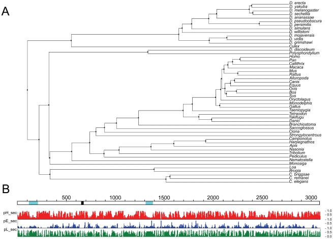
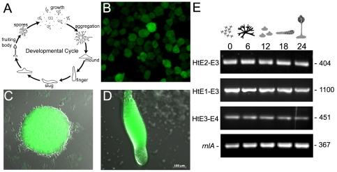
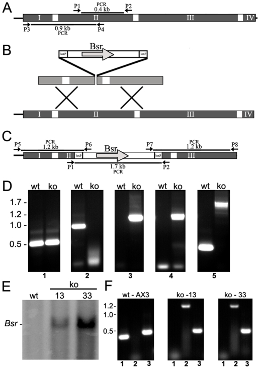
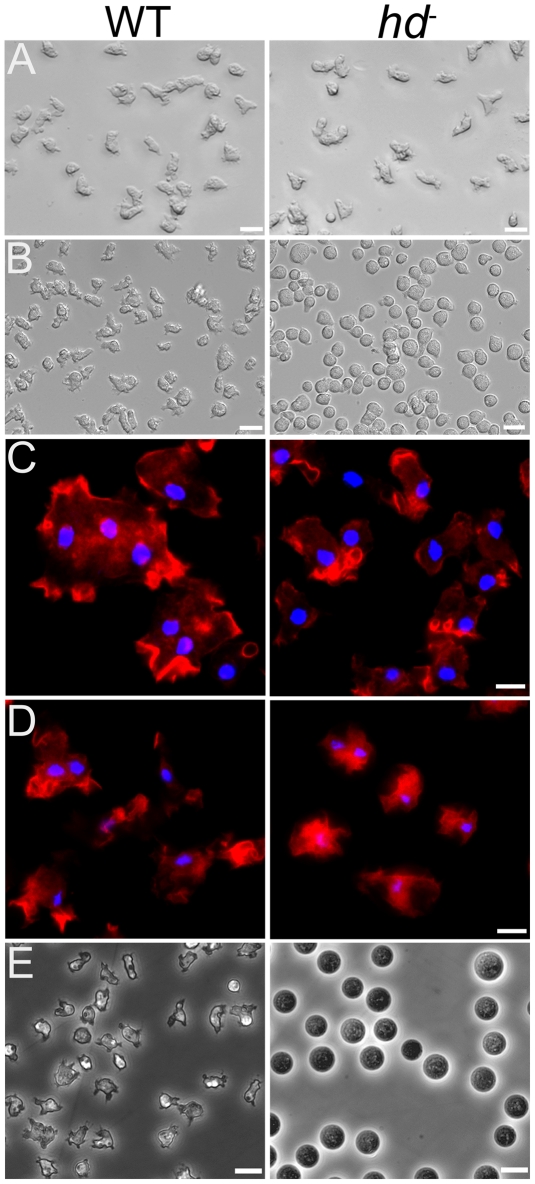
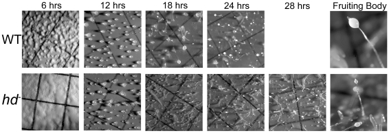
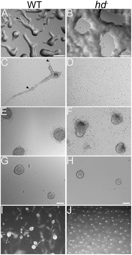
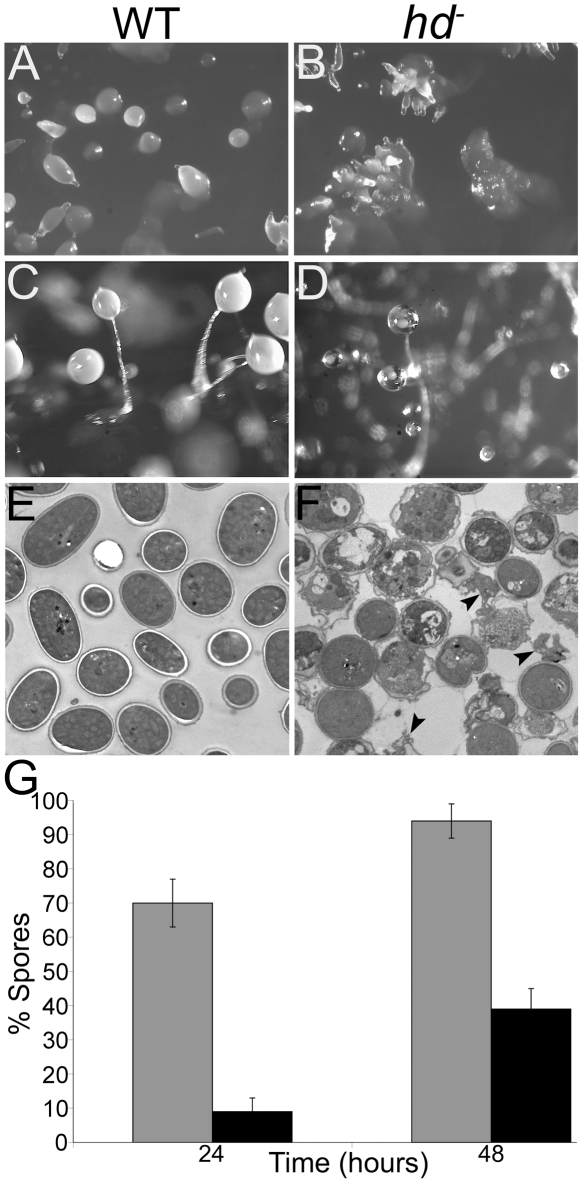
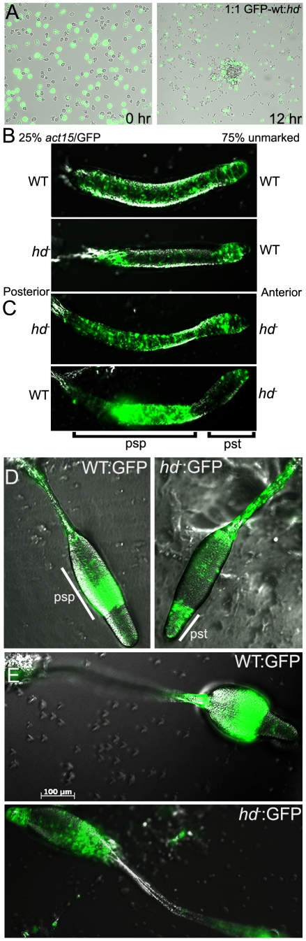
Huntingtin is a large HEAT repeat protein first identified in humans, where a polyglutamine tract expansion near the amino terminus causes a gain-of-function mechanism that leads to selective neuronal loss in Huntington's disease (HD). Genetic evidence in humans and knock-in mouse models suggests that this gain-of-function involves an increase or deregulation of some aspect of huntingtin's normal function(s), which remains poorly understood. As huntingtin shows evolutionary conservation, a powerful approach to discovering its normal biochemical role(s) is to study the effects caused by its deficiency in a model organism with a short life-cycle that comprises both cellular and multicellular developmental stages. To facilitate studies aimed at detailed knowledge of huntingtin's normal function(s), we generated a null mutant of hd, the HD ortholog in Dictyostelium discoideum. Dictyostelium cells lacking endogenous huntingtin were viable but during development did not exhibit the typical polarized morphology of Dictyostelium cells, streamed poorly to form aggregates by accretion rather than chemotaxis, showed disorganized F-actin staining, exhibited extreme sensitivity to hypoosmotic stress, and failed to form EDTA-resistant cell-cell contacts. Surprisingly, chemotactic streaming could be rescued in the presence of the bivalent cations Ca(2+) or Mg(2+) but not pulses of cAMP. Although hd(-) cells completed development, it was delayed and proceeded asynchronously, producing small fruiting bodies with round, defective spores that germinated spontaneously within a glassy sorus. When developed as chimeras with wild-type cells, hd(-) cells failed to populate the pre-spore region of the slug. In Dictyostelium, huntingtin deficiency is compatible with survival of the organism but renders cells sensitive to low osmolarity, which produces pleiotropic cell autonomous defects that affect cAMP signaling and as a consequence development. Thus, Dictyostelium provides a novel haploid organism model for genetic, cell biological, and biochemical studies to delineate the functions of the HD protein.


Conflict of interest statement

The authors have declared that no competing interests exist.

Figures

Figure 1
Figure 1. Phylogenetic and structural analysis of Dictyostelium huntingtin.
A. A phylogenetic tree showing the relationship of Dictyostelium huntingtin to huntingtin proteins from 51 other organisms was prepared as the average distance tree using BLOSUM62 of a CLUSTALW alignment in JALVIEW (reference: Waterhouse, A.M., Procter, J.B., Martin, D.M.A, Clamp, M. and Barton, G. J. (2009) “Jalview Version 2 - a multiple sequence alignment editor and analysis workbench” Bioinformatics 25 (9) 1189–1191). B. Dictyostelium huntingtin (open line) is depicted with amino acid coordinates (above) above secondary structure predictions from PROF (www.predictprotein.org; B Rost, G Yachdav and J Liu (2004) The PredictProtein Server. Nucleic Acids Research 32(Web Server issue):W321–W326) where pHsec, pEsec and pLsec represent the probability (1 high, 0 low) for helix (red), strand (blue) and neither helix nor strand (green). Locations in the huntingtin schematic of natively unstructured regions predicted by NORSnet (Avner Schlessinger and Jinfeng Liu and Burkhard Rost (2007)) and of a 19 residue polyglutamine repeat are shown by cyan and black, respectively.
Figure 2
Figure 2. The predicted endogenous hd promoter drives expression of GFP during growth and development.
(A) Developmental life cycle of Dictyostelium discoideum*. Dictyostelium spends most of its life as vegetative cells that prey upon bacteria and divide mitotically. When starved, the cells enter the developmental cycle that completes in a 24 hour period. During development, amoebae chemotactically aggregate towards secreted cAMP to form a hemispherical mound. Within the mound cells differentiate and sort out to form a motile slug. The slug ultimately forms a fruiting body comprised of a multicellular stalk supporting a ball of encapsulated dormant spores. (B) A cloned region of DNA sequence 595 bp upstream of the hd gene was used to replace the actin 15 promoter in the expression vector pTX-GFP. Wild-type AX3 cells were transformed with this construct and expression of GFP was assessed in vegetative cells; (C) mound structure showing GFP expression from hd promoter; (D) slug showing expression of GFP in both prestalk (anterior) and prespore (posterior) regions. Scale bar (µm) is shown at the bottom right. (E) Dictyostelium huntingtin is a single copy gene comprised of four exons. Total RNA was extracted from wild-type Dictyostelium strain AX3 during vegetative growth and 6-hour increments during synchronous development on nitrocellulose filters supported by filter pads soaked in DB buffer and developed at 21°C. The presence of alternate transcripts was analyzed with a combination of hd exon-specific primer sets, RT-PCR and resolved using ethidium bromide stained agarose gels. rnlA was used as a mRNA amplification control. Collection time points are shown in hours (top). hd exon boundaries are shown on the left. PCR product sizes (bp) are shown on the right. *Figure representing the developmental life cycle of Dictyostelium was adapted from CC Creative Commons Attribution – Share Alike 3.0, David Brown & Joan E. Strassmann and is freely available online for download at the dictyBase.
Figure 3
Figure 3. Targeted disruption of the hd gene by homologous recombination.
(A) Physical map of the hd gene is shown. The four exons (I, II, III and IV) that comprise the hd gene are shown (grey) interrupted by three introns (white bars). PCR primers are shown as arrows at the positions in which they prime including amplicon sizes. The exon-intron boundaries are not to drawn to scale. (B) The targeting vector and sites of recombination are shown. The vector contains the Blasticidin S resistance gene (Bsr) that is flanked by loxP sites and allows for selection of transformed cells. (C) Physical map of the targeted deletion is shown. PCR primers are shown as arrows at the positions that they prime including amplicon sizes. (D) Genomic PCR of wild-type (wt) and hd (ko) clones. Control PCR amplification of the sequence immediately upstream of the hd gene (panel 1); No amplification in clones carrying a targeted deletion of the hd gene which removes 278 bp of sequence from exon 2 (panel 2); PCR amplification of genomic DNA using primers that prime outside the vector (P5 or P8) and inside the Bsr cassette (P6 or P7), respectively (panels 3 and 4). No DNA is amplified from wild-type cells; PCR of genomic sequences that flank the insertion site (panel 5). The endogenous wild-type allele yields a fragment of ∼400 bp whereas the knockout mutant yields a much larger fragment representing insertion of the Bsr cassette. Molecular weight markers are shown on the left. (E) Confirmation of gene disruption by Southern blot analysis. Genomic DNA from wild-type or two independent hd clones (clones 13 and 33) were probed by Southern blot hybridization for the presence of Bsr sequences. (F) RT-PCR of total RNA isolated from wild-type and two independent hd clones (clones 13 and 33). In each panel, lane 1 represents the amplicon derived from priming the exon 2 deletion; lane 2 represents priming inside the Bsr cassette; and lane 3 represents control RT-PCR reactions. Molecular weight markers are shown on the left.
Figure 4
Figure 4. Removal of nutrients caused rounding of hd cells and affects F-actin localization.
(A) The morphology of hd cells (right panel) grown and photographed as an adherent culture in HL-5 was similar to wild-type cells (left panel). Scale bars (20 µm) are shown on the bottom right. (B) Removal of nutrients (HL-5) and replacement with starvation buffer (KK2) caused rounding of hd cells. Cells were photographed 20 minutes after the addition of starvation buffer. Wild-type cells (left panel) displayed morphology similar to that observed for cells in HL-5 whereas all hd cells immediately adopted a rounded morphology (right panel). Scale bars (20 µm) are shown on the bottom right. (C) Aberrant localization of F-actin in starved hd cells. Vegetative wild-type cells (left panel) and hd cells (right panel) collected from axenic medium (HL-5) were deposited into Lab-tek chambered cover glass (8 well) at 1×10⁵ cells/cm2 and allowed to grow overnight in HL-5 at 21°C. For F-actin staining, the cells were fixed with 4% formaldehyde in PDF buffer and then stained with Texas red-phalloidin. Arrows indicate membrane extensions enriched with F-actin. (D) Localization of F-actin in cells submerged in starvation buffer (KK2). One hour after starvation in KK2, wild-type cells retained their normal amoeboid morphology (left panel); hd cells remained rounded and exhibited a redistribution of F-actin away from the cell cortex as it concentrated in the cytosol (right panel). Nuclei were stained with Hoechst 33342. Scale bars (5 µm) are shown on the bottom right. (E) hd mutants were impaired in osmoregulation. Wild-type (wt) and (F) huntingtin-null (hd ) cells were grown in cell culture dishes with HL-5 media. The media was exchanged with dH2O and the cells were examined after 1 hour. Wild-type cells initially rounded and showed partial swelling under the hypoosmotic conditions, but quickly recovered the ability to crawl and change shape. In contrast, hd cells were completely round and swollen in dH2O, and barely remained attached to the culture dish. The hd cells began to rapidly lyse after ∼3 hours of this treatment and underwent complete lysis by 6 hours suggesting that the mutants may retain some very low level of osmoregulation. Scale bar 10 µm.
Figure 5
Figure 5. hd mutants exhibited asynchronous delayed development.
Exponentially growing wild-type and hd cells (2×10⁶ cells/mL) were washed three times with DB buffer, resuspended at a density of 5×10⁷ cells/mL, deposited on black nitrocellulose filters supported by filter pads soaked in DB buffer and developed for 28 hours at 21°C. All photographs are top view. Wild-type cells developed distinct aggregation territories after 6 hours whereas hd null cells displayed a much smoother appearance and the noticeable absence of streams. By 12 hours hd cells formed mounds that tended to produce multiple tips. Further development of hd cells proceeded in a delayed asynchronous manner resulting in the presence of various intermediate structures and small aborted mounds. The developmental time points in hours are shown at the top.
Figure 6
Figure 6. hd cells displayed aggregation defects and failed to stream under submerged culture.
Wild-type and hd cells (1×10⁶ cells/mL) were deposited on non-nutrient KK2 agar plates and visualized by brightfield microscopy. Images are top view. (A) Wild-type cells migrated in streams (arrows) to form aggregation territories (m) by ∼6 hours. (B) hd cells did not form well-defined streams, but rather clusters of cells that formed aggregation centers largely by accretion. Scale bar 100 µm. (C) Wild-type and (D) hd cells (1×10⁵ cells/cm2) were submerged under KK2 and allowed to develop for 6 hours. Streams (arrows) of wild-type cells moving into an aggregation center (m) were readily apparent (left panel) whereas hd cells failed to stream (right panel). (E) Wild-type and (F) hd cells were submerged under buffer at high density (5×10⁶ cells/cm2) and imaged after 20 hours. Under these conditions, wild-type cells readily formed distinct organized mounds; high density partially restored the ability of hd cells to form aggregation territories but with an irregular polarized shape. (G) Wild-type and (H) hd cells (1×10⁵ cells/cm2) were submerged under KK2 in the presence of 1 mM CaCl2 and imaged after 20 hours. Under these conditions exogenous calcium rescues early development of hd cells. Scale bar 100 µm. (I) Development on KK2 agar in the presence of EGTA. After 24 hours, wild type cells form fruiting bodies whereas development of hd cells (J) is blocked in the presence of 1 mM EGTA.
Figure 7
Figure 7. Loss of EDTA-resistant adhesion in hd cells.
Wild-type and hd cells (5×10⁶ cells/mL) were developed in Soerensen's buffer at 150 rpm and 21°C. Samples were collected at the start of the assay and at one hour time points over a period of 6 hours. For each collection point, cells were dissociated by vortexing and then incubated in the presence or absence of 10 mM EDTA for 30 minutes, fixed with 2% glutaraldehyde (10 min.) and single cells were counted in triplicate using a hemocytometer. All experiments were performed in duplicate at least three times and the mean value for single cells in duplicate samples, expressed as percentage of total cells was plotted over time.
Figure 8
Figure 8. hd cells formed mounds with multiple prestalk-tips and fruiting bodies with a glassy sorus.
(A) Wild-type mounds formed mounds that develop a single tip which proceeded to form an elongated finger structure. (B) Multiple prestalk-tips formed atop most hd mounds that went on to form comparably small individual finger structures. Cells were developed on SM agar with a lawn of Klebsiella. Images are top view. (C) Terminal structures of wild-type (left panel) and (D) hd null cells (right panel). After 36–48 hours, the sori of hd cells became progressively glassy in appearance and contained very few spores. (E) hd spores spontaneously germinated within the sorus. Wild-type and hd spores were collected and fixed (see Materials and Methods) for imaging using electron microscopy. Wild-type sori contained intact elliptical dormant spores with well-defined spore coats and without the presence of amoebae. (F) Spores collected from hd fruiting bodies showed a heterogeneous mixture of round spores that represented the various stages of germination. Swollen spores, swollen spores with thin spore walls about to release amoebae, and nascent amoebae (arrows) are shown. (G) hd mutants displayed defects in prespore/spore cell differentiation. Wild-type and hd cells were starved at low density in monolayer culture in the presence of 15 mM 8-Br-cAMP to induce sporulation, viewed by brightfield microscopy and the number of spores formed was counted. The percentage of differentiated wild-type (grey bars) and hd (black bars) spore cells was calculated from the total cells after 24 and 48 hour incubation periods. On average, hd cells collectively formed spore cells at ∼30% efficiency relative to wild-type controls after 48 hours. Bars indicate standard errors that are derived from three independent experiments, each with three replicates.
Figure 9
Figure 9. hd cells produced spores with reduced viability.
The viability of wild-type (grey bars) and hd (black bars) spores was assessed. Spores were untreated, heated to 45°C for 10 minutes or incubated with 0.5% NP40 detergent for 5 minutes and aliquots of 100 spores were plated in triplicate onto SM-5 agar plates in a suspension of bacteria and grown for 7 days at 21°C. The relative viability of hd spores was assessed by counting the number of clear plaques formed on the bacterial lawns. Results are representative of three independent experiments.
Figure 10
Figure 10. Huntingtin regulates prespore/spore differentiation cell-autonomously.
GFP was expressed in wild-type cells and their position in chimeric aggregation territories with unlabelled hd cells was monitored. (A) Mixed (1∶1) cell suspensions were submerged (1×10⁵ cells/cm2) under KK2 buffer (left panel). After 12 hours, aggregation centers were imaged under brightfield and fluorescence using an inverted microscope. hd cells failed to populate the central region of aggregation territories but were instead localized to the periphery of the mound. Images are representative of three independent experiments. (B) Wild-type and hd cells that express GFP were mixed 1∶3 or less with unmarked wild-type cells as indicated and cellular organization in live cells during the slug stage was assessed. GFP-labeled wild-type cells were mixed with the same genotype as a control (top panel); GFP-labeled hd cells populated the prestalk regions of the slug (lower panel). (C) GFP-labeled hd cells were mixed with the same genotype as a control (top panel); GFP-labeled wild-type cells populated the prespore regions of the slug (lower panel). Slugs are positioned with their indicated posterior prespore (psp) zones to the left and anterior prestalk (pst) zones to the right. (D) hd cells marked with an actin/GFP reporter plasmid were mixed with unmarked wild-type cells in a 1∶3 ratio and allowed to develop into fruiting bodies. Developing structures (late culminant) were imaged by DIC and fluorescent microscopy. Wild-type cells marked with GFP (left panel) predominantly occupied the prespore/spore region; hd cells marked with GFP failed to populate the prespore region and were overrepresented in the upper cup, lower cup and basal disc (right panel). Psp – prespore; Pst – prestalk. (E) Terminal fruiting structures were viewed by DIC and fluorescent microscopy. Wild-type cells expressing GFP (top panel); hd cells expressing GFP (lower panel). Results are representative of three independent experiments for all panels shown. Scale bar 100 µm.

Similar articles

Cited by

References

    1. Vonsattel JP, Myers RH, Stevens TJ, Ferrante RJ, Bird ED, et al. Neuropathological classification of Huntington's disease. J Neuropathol Exp Neurol. 1985;44(6):559–577. - PubMed
    1. Martin JB, Gusella JF. Huntington's disease: Pathogenesis and management. N Engl J Med. 1986;315(20):1267–1276. - PubMed
    1. Gusella JF, Wexler NS, Conneally PM, Naylor SL, Anderson MA, et al. A polymorphic DNA marker genetically linked to Huntington's disease. Nature. 1983;306(5940):234–238. - PubMed
    1. Gusella JF, MacDonald ME. Huntington's Disease. Semin Cell Biol. 1995;6:21–28. - PubMed
    1. The Huntington's Disease Collaborative Research Group. A novel gene containing a trinucleotide repeat that is expanded and unstable on Huntington's Disease chromosomes. Cell. 1993;72:971–983. - PubMed

Publication types

MeSH terms