

doi: 10.1002/adma.200501726.
Conducting-Polymer Nanotubes for Controlled Drug Release
Affiliations
- PMID: 21552389
- PMCID: PMC3088882
- DOI: 10.1002/adma.200501726
Item in Clipboard
Conducting-Polymer Nanotubes for Controlled Drug Release
Adv Mater.
.
No abstract available
Figures
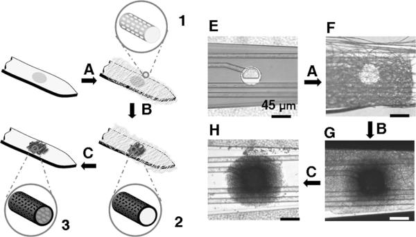
Schematic diagrams illustrating the surface modification of neural microelectrodes to create nanotubular PEDOT: A) electrospinning of biodegradable polymer (PLGA) fibers with well-defined surface texture (1) on the probe tip, B) electrochemical polymerization of conducting polymers (PEDOT) (2) around the electrospun fibers, and C) dissolving the electrospun core fibers to create nanotubular conducting polymers (3). Optical micrograph of: E) the gold electrode site, F) the electrode site after electrospinning showing the coverage of the PLGA electrospun nanoscale fibers, G) the electrode after electrochemical deposition of PEDOT on the gold site and around the electrospun fibers, and H) the electrode after removal of the core nanoscale fiber templates.
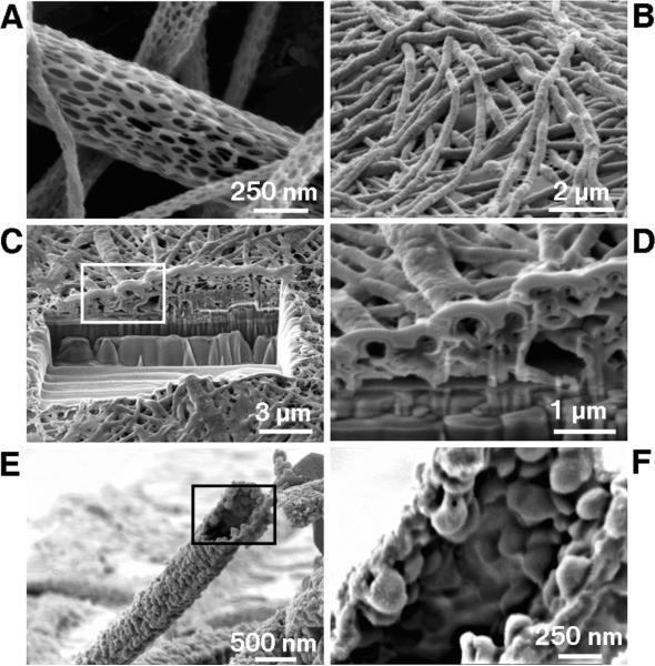
Scanning electron micrographs of PLGA nanoscale fibers and PEDOT nanotubes. A) diameters of the PLGA fibers were distributed over the range 40–500 nm with the majority being between 100–200 nm. B) Electropolymerized PEDOT nanotubes on the electrode site of an acute neural probe tip after removing the PLGA core fibers. C) A section of (B) cut with a FIB showing the silicon substrate layer and PEDOT nanoscale fiber coating. D) Higher-magnification image of (C) showing the PEDOT nanotubes crossing each other. E) A single PEDOT nanotube which was polymerized around a PLGA nanoscale fiber, followed by dissolution of the PLGA core fiber. This image shows the external texture at the surface of the nanotube. F) Higher-magnification image of a single PEDOT nanotube demonstrating the textured morphology that has been directly replicated from the external surface of the electrospun PLGA fiber templates. The average wall thickness of PEDOT nanotubes varied from 50–100 nm, with the nanotube diameters ranging from 100 to 600 nm.
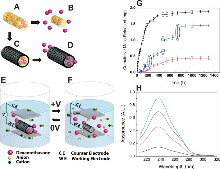
Schematic illustration of the controlled release of dexamethasone: A) dexamethasone-loaded electrospun PLGA, B) hydrolytic degradation of PLGA fibers leading to release of the drug, and C) electrochemical deposition of PEDOT around the dexamethasone-loaded electrospun PLGA fiber slows down the release of dexamethasone (D). E) PEDOT nanotubes in a neutral electrical condition. F) External electrical stimulation controls the release of dexamethasone from the PEDOT nanotubes due to contraction or expansion of the PEDOT. By applying a positive voltage, electrons are injected into the chains and positive charges in the polymer chains are compensated. To maintain overall charge neutrality, counterions are expelled towards the solution and the nanotubes contract. This shrinkage causes the drugs to come out of the ends of tubes. G) Cumulative mass release of dexamethasone from: PLGA nanoscale fibers (black squares), PEDOT-coated PLGA nanoscale fibers (red circles) without electrical stimulation, and PEDOT-coated PLGA nanoscale fibers with electrical stimulation of 1 V applied at the five specific times indicated by the circled data points (blue triangles). H) UV absorption of dexamethasone-loaded PEDOT nanotubes after 16 h (black), 87 h (red), 160 h (blue), and 730 h (green). The UV spectra of dexamethasone have peaks at a wavelength of 237 nm. Data are shown with a ± standard deviation (n = 15 for each case).
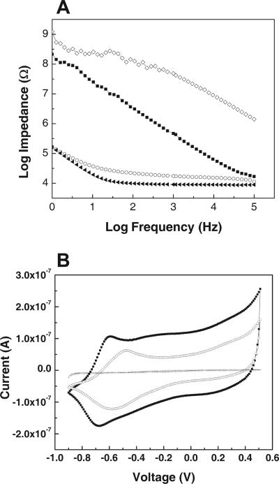
Impedance spectroscopy and CV: A) impedance spectrum of an acute neural probe site over a frequency range of 1–105 kHz: bare gold (filled squares), with electrospun PLGA fiber templates (diamonds), with PLGA fibers and electrochemical polymerized PEDOT (circles), or with PEDOT nanotubes by removing the PLGA core fibers (filled triangles). B) CV of an acute neural probe: bare gold (crosses), with PLGA electrospun fiber templates and electrochemical deposition of PEDOT (circles), or with PEDOT nanotube fibers prepared by removing the PLGA core fibers (filled squares). The potential was swept from –0.9 to 0.5 V at a scan rate of 100 mV s–1.
References
-
- Devoret MH, Esteve D, Urbina C. Nature. 1992;360:547.
-
- Ozin GA. Adv. Mater. 1992;4:612.
-
- Gref R, Minamitake Y, Peracchia MT, Trubetskoy V, Torchilin V, Langer R. Science. 1994;263:1600. - PubMed
-
- Parthasarathy RV, Martin CR. Nature. 1994;369:298. - PubMed
-
- Whitesides GM, Mathias JP, Seto CT. Science. 1991;254:1312. - PubMed
Grants and funding
LinkOut - more resources
Full Text Sources
Other Literature Sources
