. 2011;2(2):111-8.

A new crystal form of mouse thiamin pyrophosphokinase

Affiliations

A new crystal form of mouse thiamin pyrophosphokinase

Jing-Yuan Liu et al. Int J Biochem Mol Biol. 2011.

Abstract

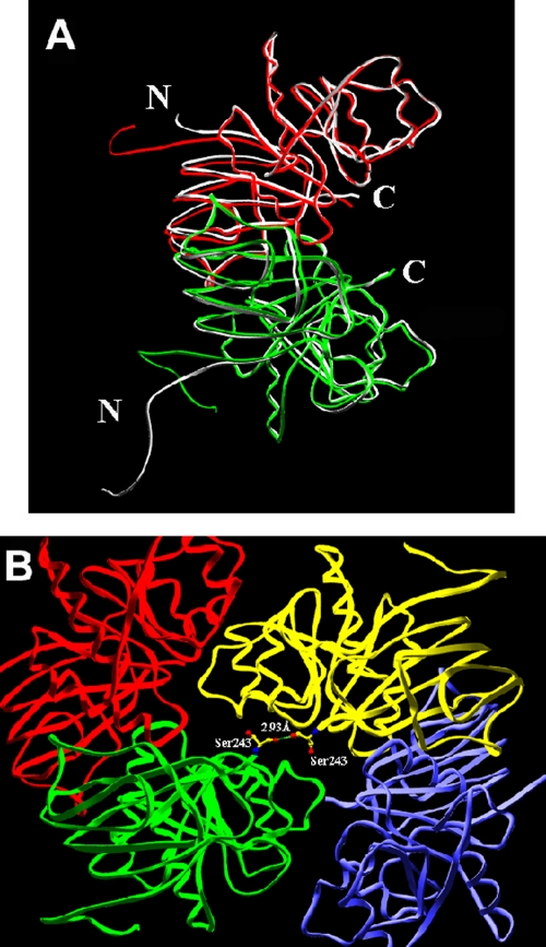
Thiamin pyrophosphokinase (TPK) transfers a pyrophosphate group from ATP to the hydroxyl group of thiamin and produces thiamin pyrophosphate (TPP). TPP is the cofactor of metabolically important enzymes such as pyruvate dehydrogenase, α-ketoglutarate dehydrogenase, branched-chain α-keto acid dehydrogenase, transketolase and 2-hydroxyphytanoyl-CoA lyase. Thiamin deficiency results in Wernike-Korsakof Syndrome (WKS) due to neurological disorder and wet beriberi, a potentially fatal cardiovascular disease. Mouse TPK associates as a dimer revealed by previous solved crystallographic structures. In this study, we report mouse TPK complexed with TPP-Mg(2+) and thiamin -Mg(2+), respectively, in a new crystal form. In these two structures, four mouse TPK molecules were found in each asymmetric unit. Although we cannot rule out this tetramer form can be an artifact from crystal packing, mouse TPK tetramer has a more closed ATP binding pocket and has the potential to provide specific interactions between mouse TPK and ATP compared with the previous dimeric structure and is likely to be an active form.

Keywords: TPP-Mg2+; Thiamin pyrophosphokinase (TPK); crystal structure; dimer; protein oligomerization; tetramer; thiamin-Mg2+.


Figures

Figure 1.
Figure 1.
The overall structure of mouse TPK tetramer. A. A view of the tetramer structure parallel to its two fold axis. B. A view of the tetramer structure perpendicular to its two fold axis. The four subunits assigned as A, B, C and D are colored in green, red, yellow and blue, respectively. A and B or C and D form the previously described U-shaped dimer unit.
Figure 2.
Figure 2.
Differences between a dimer unit of the tetrameric structure and the dimeric structure of mouse TPK. A. Superposition of mouse TPK dimer (in gray) and tetramer (in green and red). The N-terminal His tags are oriented differently and the C-termini are pointed to different directions compared with the dimeric structure. B. A zoom in picture of the two C-terminal Ser in A and C subunits. The arrangsement of the tetramer and the shift of the C-terminal residue, Ser243, of A and C subunit brought the two residues close enough to form a hydrogen bond between the hydroxyl oxygen of Ser in A subunit and the carbonyl oxygen of Ser in C subunit.
Figure 3.
Figure 3.
Molecular surface of the dimeric and tetrameric structures. A. Molecular surface of the dimeric ternary complex of mouse TPK with PPP-MgAMP. AMP is shown in orange. PPP is shown in green. The binding groove is open to the solvent. B. The tetramer mouse TPK is superimposed with the dimer structure and the molecular surface is shown. The joining of another dimer unit helps to seal the active site. C. Glu57 may provide additional hydrogen bond across the two dimer interface in the tetrameric structure when nucleotide is bound. The dimeric PPP -MgAMP mouse TPK complex is superimposed onto the tetrameric structure. PPP and Mg2+ are shown in magenta. AMP is shown in red. Distance is shown in angstrom. The dimer structure is shown in yellow; the A and C subunits of the I tetramer structure are shown in blue and green. The common I part of the two structures is illustrated by ribbon diagram. The C subunit of tetramer, which does not exist in the dimer structure, is in stick presentation. Glu57 in tetramer is 4.96 Å away from the N6 atom of AMP in the dimer structure.
Figure 4.
Figure 4.
Magnesium binding in tetrameric TPP-Mg and thiamin-Mg structure complex. A. Magnesium binding in TPP-Mg mouse TPK complex. Magnesium coordinates with the α-phosphate of TPP and Asp 46, 71 and 73. B. Superposition of thiamin molecules and residues surrounding magnesium ion of the dimeric (in green) and tetrameric (in red) structure. Magnesium ion binds to the enzyme via coordination with Asp 46, 71, 73 and 100. The hydroxyl group of thiamin shifted and the hydrogen bond, which used to exist between the hydroxyl group of thiamin and Asp71 in the dimeric structure, is abolished in the tetrameric structure. Distance is shown in angstrom.

Similar articles

Cited by

References

    1. Ishii K, Sarai K, Sanemori H, Kawasaki T. Concentrations of thiamine and its phosphate esters in rat tissues determined by high-performance liquid chromatography. J Nutr SciVitaminol (Tokyo) 1979;25:517–523. - PubMed
    1. Kimura M, Itokawa Y. Determination of thiamin and thiamin phosphate esters in blood by liquid chromatography with post-column derivatization. Clin Chem. 1983;29:2073–2075. - PubMed
    1. Bettendorff L, Peeters M, Wins P, Schoffeniels E. Metabolism of thiamine triphosphate in rat brain: correlation with chloride permeability. J Neurochem. 1993;60:423–434. - PubMed
    1. Shikata H, Egi Y, Koyama S, Yamada K, Kawasaki T. Properties of the thiamin triphosphate-synthesizing activity catalyzed by adenylate kinase (isoenzyme 1) Biochem Int. 1989;18:943–949. - PubMed
    1. Nosaka K, Kaneko Y, Nishimura H, Iwa-shima A. A possible role for acid phosphatase with thiamin-binding activity encoded by PH03 in yeast. FEMS Microbiol Lett. 1989;51:55–59. - PubMed

LinkOut - more resources