

Novel TSPAN12 mutations in patients with familial exudative vitreoretinopathy and their associated phenotypes
- PMID: 21552475
- PMCID: PMC3087453
Novel TSPAN12 mutations in patients with familial exudative vitreoretinopathy and their associated phenotypes
Abstract
Purpose: Mutations in tetraspanin 12 (TSPAN12) have recently been identified as a cause of autosomal dominant familial exudative vitreoretinopathy (FEVR). The purpose of this study was to detect TSPAN12 mutations in Chinese patients with FEVR and to describe the associated phenotypes.
Methods: Sanger sequencing was used to analyze the seven coding exons and their adjacent regions of TSPAN12 in 49 unrelated FEVR patients. Clinical phenotypes of the patients with TSPAN12 mutations were documented.
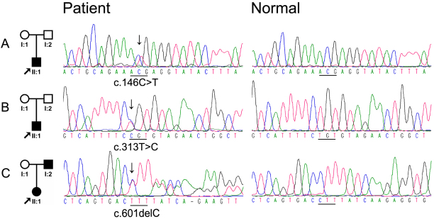
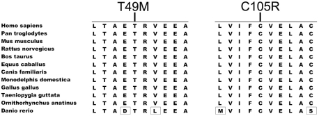
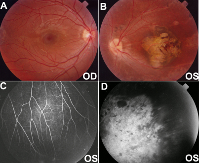
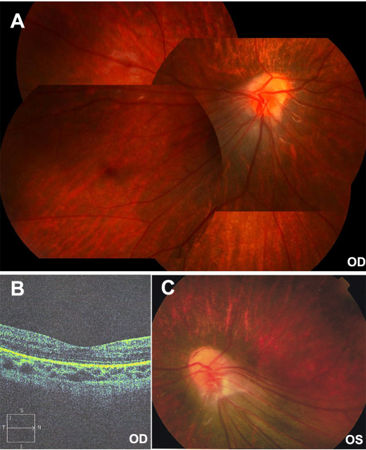
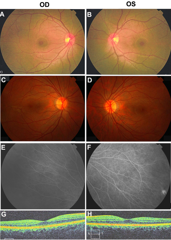
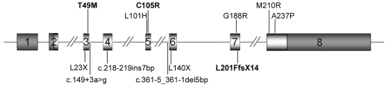
Results: Three novel heterozygous mutations in TSPAN12 were identified in three patients from unrelated families: c.146C>T (p.Thr49Met), c.313T>C (p.Cys105Arg), and c.601delC (p.Leu201PhefsX14). All three mutations involved highly conserved residues and were not present in 180 normal individuals. Ocular phenotypes included retinal folds, inferotemporal dragging of the optic disc and macula, increased vessels in the equatorial region, and a peripheral avascular zone. A father and his daughter had the same mutation but the father only had mild peripheral fundus changes while his daughter had obvious dragged disc and macular ectopia.
Conclusions: Our results suggest that TSPAN12 mutations are responsible for FEVR. Similar to patients with mutations in NDP, LRP5, or FZD4, the phenotypes associated with TSPAN12 mutations showed great variations between different individuals within a family and between the two eyes in individual patients.
Figures
Similar articles
-
Novel mutations in the TSPAN12 gene in Chinese patients with familial exudative vitreoretinopathy.Mol Vis. 2014 Sep 20;20:1296-306. eCollection 2014. Mol Vis. 2014. PMID: 25352738 Free PMC article.
-
Mutation Spectrum of the LRP5, NDP, and TSPAN12 Genes in Chinese Patients With Familial Exudative Vitreoretinopathy.Invest Ophthalmol Vis Sci. 2017 Nov 1;58(13):5949-5957. doi: 10.1167/iovs.17-22577. Invest Ophthalmol Vis Sci. 2017. PMID: 29181528
-
Identification of LRP5 mutations in families with familial exudative vitreoretinopathy.Yi Chuan. 2017 Mar 20;39(3):241-249. doi: 10.16288/j.yczz.16-339. Yi Chuan. 2017. PMID: 28420620
-
Novel Exon 7 Deletions in TSPAN12 in a Three-Generation FEVR Family: A Case Report and Literature Review.Genes (Basel). 2023 Feb 25;14(3):587. doi: 10.3390/genes14030587. Genes (Basel). 2023. PMID: 36980859 Free PMC article. Review.
-
Familial exudative vitreoretinopathy and related retinopathies.Eye (Lond). 2015 Jan;29(1):1-14. doi: 10.1038/eye.2014.70. Epub 2014 Oct 17. Eye (Lond). 2015. PMID: 25323851 Free PMC article. Review.
Cited by
-
Pathogenic variants and associated phenotypic spectrum of TSPAN12 based on data from a large cohort.Graefes Arch Clin Exp Ophthalmol. 2021 Oct;259(10):2929-2939. doi: 10.1007/s00417-021-05196-y. Epub 2021 Apr 27. Graefes Arch Clin Exp Ophthalmol. 2021. PMID: 33907885
-
Mutation spectrum of the FZD-4, TSPAN12 AND ZNF408 genes in Indian FEVR patients.BMC Ophthalmol. 2016 Jun 17;16:90. doi: 10.1186/s12886-016-0236-y. BMC Ophthalmol. 2016. PMID: 27316669 Free PMC article.
-
Migrasome and Tetraspanins in Vascular Homeostasis: Concept, Present, and Future.Front Cell Dev Biol. 2020 Jun 16;8:438. doi: 10.3389/fcell.2020.00438. eCollection 2020. Front Cell Dev Biol. 2020. PMID: 32612990 Free PMC article. Review.
-
Familial Exudative Vitreoretinopathy-Related Disease-Causing Genes and Norrin/β-Catenin Signal Pathway: Structure, Function, and Mutation Spectrums.J Ophthalmol. 2019 Nov 16;2019:5782536. doi: 10.1155/2019/5782536. eCollection 2019. J Ophthalmol. 2019. PMID: 31827910 Free PMC article. Review.
-
Identification and Characterization of ATOH7-Regulated Target Genes and Pathways in Human Neuroretinal Development.Cells. 2024 Jul 3;13(13):1142. doi: 10.3390/cells13131142. Cells. 2024. PMID: 38994994 Free PMC article.
References
-
- Criswick VG, Schepens CL. Familial exudative vitreoretinopathy. Am J Ophthalmol. 1969;68:578–94. - PubMed
-
- van Nouhuys CE. Dominant exudative vitreoretinopathy. Ophthalmic Paediatr Genet. 1985;5:31–8. - PubMed
-
- Feldman EL, Norris JL, Cleasby GW. Autosomal dominant exudative vitreoretinopathy. Arch Ophthalmol. 1983;101:1532–5. - PubMed
-
- Gow J, Oliver GL. Familial exudative vitreoretinopathy. An expanded view. Arch Ophthalmol. 1971;86:150–5. - PubMed
Publication types
MeSH terms
Substances
Supplementary concepts
LinkOut - more resources
Full Text Sources
Other Literature Sources
Medical