. 2011 Apr 29:17:1110-27.

Degeneration of the mouse retina upon dysregulated activity of serum response factor

Affiliations

Degeneration of the mouse retina upon dysregulated activity of serum response factor

Jenny Sandström et al. Mol Vis. .

Abstract

Purpose: Our aim was to generate and phenotypically characterize a transgenic mouse line expressing a constitutively active variant of the transcription regulatory protein serum response factor (SRF), namely the SRF-VP16 protein. This new mouse strain has been registered under the designation Gt(ROSA)26Sor(tm1(SRF-VP16)Antu). We found phenotypic changes upon ectopic expression of SRF-VP16, especially in the mouse retina.

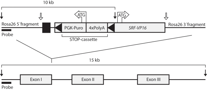
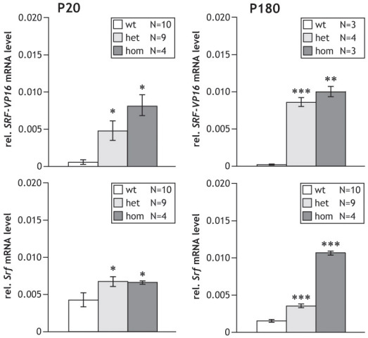
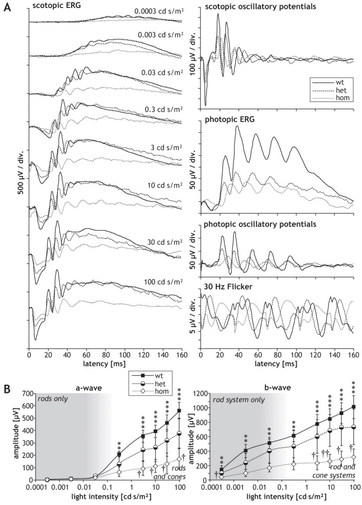
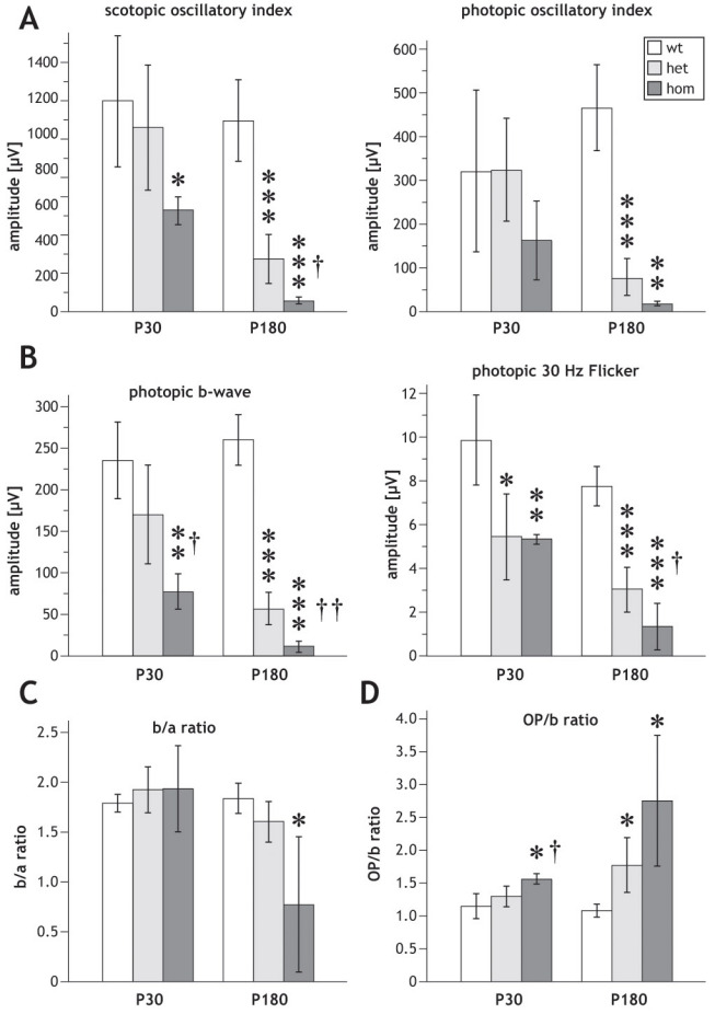
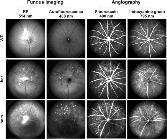
Methods: Using homologous recombination, we integrated an SRF-VP16 conditional (i.e., "flox-STOP" repressed) expression transgene into the Rosa26 locus of murine embryonic stem (ES) cells. These engineered ES cells were used to derive the Gt(ROSA)26Sor(tm1(SRF-VP16)Antu) mouse strain. Semiquantitative real-time PCR was used to determine expression of the SRF-VP16 transgene at the mRNA level, both in young (P20 and P30) and adult (six months old) Gt(ROSA)26Sor(tm1(SRF-VP16)Antu) mice. We also investigated the transcript levels of endogenous Srf and several SRF target genes. Retinal function was tested by electroretinography in both young and adult mice. Morphological abnormalities could be visualized by hematoxylin and eosin staining of sectioned, paraffin-embedded eye tissue samples. Scanning-laser ophthalmoscopy was used to investigate retinal vascularization and degeneration in adult mice.

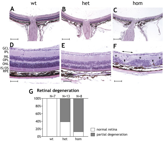
Results: We show that the SRF-VP16 mRNA is expressed to a low but significant degree in the retinas of young and adult animals of the Gt(ROSA)26Sor(tm1(SRF-VP16)Antu) mouse strain, even in the absence of Cre-mediated deletion of the "flox-STOP" cassette. In the retinas of these transgenic mice, endogenous Srf displays elevated transcript levels. Ectopic retinal expression of constitutively active SRF-VP16 is correlated with the malfunction of retinal neurons in both heterozygous and homozygous animals of both age groups (P20 and adult). Additionally, mislamination of retinal cell layers and cellular rosette formations are found in retinas of both heterozygous and homozygous animals of young age. In homozygous individuals, however, the cellular rosettes are more widespread over the fundus. At adult age, retinas both from animals that are heterozygous and homozygous for the floxSTOP/SRF-VP16 transgene display severe degeneration, mainly of the photoreceptor cell layer. Wild-type age-matched littermates, however, do not show any degeneration. The severity of the observed effects correlates with dosage of the transgene.

Conclusions: This is the first report suggesting an influence of the transcription factor SRF on the development and function of the murine retina. Ectopic SRF-VP16 mRNA expression in the retinas of young animals is correlated with photoreceptor layer mislamination and impaired retinal function. At an advanced age of six months, degenerative processes are detected in SRF-VP16 transgenic retinas accompanied by impaired retinal function. The Gt(ROSA)26Sor(tm1(SRF-VP16)Antu) mouse strain represents a genetic SRF gain-of-function mouse model that will complement the current SRF loss-of-function models. It promises to provide new insight into the hitherto poorly defined role of SRF in retinal development and function, including potential contributions to ophthalmologic disorders. Furthermore, using conditional Cre-mediated activation of SRF-VP16, the described mouse strain will enable assessment of the impact of dysregulated SRF activity on the physiologic functions of various other organs.


Figures

Figure 1
Figure 1
Schematic representation of the Rosa26-floxSTOP/SRF-VP16 DNA targeting construct. The Rosa26-floxSTOP/SRF-VP16 DNA targeting construct (upper) and its integration site in the wild-type genomic Rosa26 locus (lower) are shown. Black arrows indicate EcoRI restriction sites, applied to Southern blot detection of the targeting construct (10 kb band) and the wild-type ROSA26 locus (15 kb band), using the probe indicated. White arrows indicate XbaI restriction sites generated upon the insertion of the floxSTOP/SRF-VP16 construct into the ROSA26–1 vector. The black box depicted upstream of the Puro-PGK cassette represents the splice acceptor. Black triangles indicate the two loxP sites flanking the STOP cassette.
Figure 2
Figure 2
Retinal mRNA levels of SRF-VP16 and endogenous Srf at ages P20 and P180. Real-time PCR quantification of relative SRF-VP16 and endogenous Srf mRNA levels in the retinas of P20 and P180 mice. SRF-VP16 RNA levels were only observed in retinas of Rosa26-floxSTOP/SRF-VP16 transgenic mice, both heterozygous (het) and homozygous (hom) for the SRF-VP16 transgene (upper panels). Expression of endogenous Srf was always elevated compared to wild-type mice in the presence of the transgene; this was especially pronounced in the transgenic P180 (hom) mice (lower panels). Error bars indicate SEM, *p<0.05, **p<0.01, and ***p<0.001, denoting significance as compared to wild-type mice. All transcriptional levels were normalized to Gapdh.
Figure 3
Figure 3
Graphs presenting electroretinographic measurements of animals at age P30. Electoretinographic (ERG) waveforms obtained in P30 (one-month-old) mice of the Rosa26 (wt), Rosa26-floxSTOP/SRF-VP16 (het), and Rosa26-floxSTOP/SRF-VP16 (hom) genotypes. A: Typical waveforms are shown of scotopic and photopic ERGs, scotopic and oscillatory potentials, as well as the photopic 30 Hz flicker, as indicated. Note the different scaling. B: Values of amplitudes of scotopic a-waves and b-waves, depending on the intensity of light stimuli. Significances of differences between wild-type and homozygous mice are indicated by asterisks, and significances of the differences between heterozygous and homozygous mice is indicated by crosses (* or †p<0.05, ** or ††p<0.01, ***p<0.001).
Figure 4
Figure 4
Quantification of electroretinographic parameters. Comparison of several electroretinographic (ERG) parameters obtained in P30 and P180 Rosa26 wild-type (wt), Rosa26-floxSTOP/SRF-VP16 (het), and Rosa26-floxSTOP/SRF-VP16 (hom) mice, respectively, as indicated. A: Scotopic- (left) and photopic (right) oscillatory index. B: Photopic b-wave (left) and photopic 30Hz flicker (right). C: b/a ratio. D: OP/b ratio. Significances of differences between wild-type and transgenic mice are indicated by asterisks, and significances of the differences between heterozygous and homozygous mice are indicated by crosses (* or †p<0.05, ** or ††p<0.01, ***p<0.001).
Figure 5
Figure 5
Graphs presenting electroretinographic measurements of animals at age P180. Electroretinographic (ERG) measurements obtained with P180 (six-month-old) mice of the Rosa26 (wt), Rosa26-floxSTOP/SRF-VP16 (het), and Rosa26-floxSTOP/SRF-VP16 (hom) genotypes. A: Typical waveforms are shown of scotopic and photopic ERGs, scotopic and oscillatory potentials, as well as the photopic 30 Hz flicker, as indicated. Note the different scaling. B: Values of amplitudes of scotopic a-waves and b-waves depending on the intensity of light stimuli are shown. Amplitudes measured in wild-type mice are larger than amplitudes obtained in the mutants, with high significance over almost all light intensities (***p<0.001).
Figure 6
Figure 6
Rosette formations in the photoreceptor layer of P20 animals heterozygous or homozygous for the Rosa26-floxSTOP/SRF-VP16 transgene. Histological sections are provided displaying rosette formations in the photoreceptor layers of Rosa26- floxSTOP/SRF-VP16 heterozygous or homozygous P20 animals. A-C: Hematoxylin and eosin–stained sections are shown of retinas obtained from wild-type, heterozygous, and homozygous P20 animals. Scale bar represents 200 μm. A: Normal wild-type retina is displayed and compared to those of heterozygous (B) and homozygous (C) transgenic animals, showing the localization of rosette formation around the vicinity of the optic nerve. D-F: Magnifications are shown of regions of the retinas displaying normal (D) or dyslaminated (E, F) retinal layers. Normal mislamination in wild-type retinas (D) with the ganglion cell layer (GCL), inner plexiform layer (IPL), inner nuclear layer (INL), outer nuclear layer (ONL), inner and outer segments (IS/OS), and retinal pigment epithelium (RPE). Rosette formations to a varying extents are shown in heterozygous (E) and homozygous (F) P20 retinas. Scale bars represent 50 μm. G: Quantification of rosette formation is shown. Retinas displaying rosette formations were categorized as positive for rosette formation. Eighty-seven percent of the heterozygous animals showed rosette formation, while 100% homozygous animals displayed rosette structures.
Figure 7
Figure 7
Retinal degeneration in P180 mice heterozygous or homozygous for the Rosa26-floxSTOP/SRF-VP16 transgene. Histological sections of P180 retinas are shown displaying degenerative processes in Rosa26-floxSTOP/SRF-VP16 heterozygous (het) and homozygous (hom) animals. A-C: Hematoxylin and eosin staining. Note the scattered degeneration in the posterior part of heterozygous and homozygous retinas. Peripheral regions show no degeneration. Scale bars represent 200 μM. D-F: Higher magnifications of P180 retinas are displayed. Wild-type retina (D) showing ganglion cell layer, inner plexiform layer, inner nuclear layer, outer nuclear layer, inner and outer segments and retinal pigment epithelium (RPE). Degeneration of the photoreceptor layer in adult heterozygous (E) and homozygous (F) retinas is shown. In (F), arrowheads point to regions of complete degeneration of the inner and outer nuclear layers, whereas inner nuclear layers can be found at least partially in other places (asterisks). Occasionally, even the ganglion cell layer can no longer be found (indicated by horizontal double-headed arrow). Scale bars represent 50 μM. G: Quantification of individual mice displaying retinal degeneration in histological sections. Retinas displaying areas in the outer nuclear layer with eight or fewer layers of photoreceptor nuclei were classified as having “partial retinal degeneration.” Sixty percent of the heterozygous animals and over 80% of the homozygous animals showed partial degeneration.
Figure 8
Figure 8
Scanning-laser ophthalmoscopy (SLO) imaging. Eyes from Rosa26 (wt; upper row), Rosa26-floxSTOP/SRF-VP16 (het) (middle row), and Rosa26-floxSTOP/SRF-VP16 (hom; bottom row) animals at six months of age (P180) were investigated. Fundus imaging with RF at 514 nm excitation (left column) and autofluorescence at 488 nm laser wavelengths (middle left column) display dramatic changes in fundus appearance, structure of the nerve fiber layer and accumulation of autofluorescent lipids in genetically modified animals compared to wild-type animals. Retinal degenerative processes can also be inferred from enhanced angiographic deep layer (choroidal) signals using fluorescein (middle right column) and indocyanine green (right column) angiography, as clearly detected in both heterozygous and homozygous animals.

Similar articles

Cited by

References

    1. Graw J. Genetic aspects of embryonic eye development in vertebrates. Dev Genet. 1996;18:181–97. - PubMed
    1. Jean D, Ewan K, Gruss P. Molecular regulators involved in vertebrate eye development. Mech Dev. 1998;76:3–18. - PubMed
    1. Rossant J, Tam PPL, editors. Mouse development: Patterning, morphogenesis, and organogenesis. 1st edition. San Diego: Academic Press; 2002.
    1. Alberti S, Krause SM, Kretz O, Philippar U, Lemberger T, Casanova E, Wiebel FF, Schwarz H, Frotscher M, Schütz G, Nordheim A. Neuronal migration in the murine rostral migratory stream requires serum response factor. Proc Natl Acad Sci USA. 2005;102:6148–53. - PMC - PubMed
    1. Etkin A, Alarcón JM, Weisberg SP, Touzani K, Huang YY, Nordheim A, Kandel ER. A role in learning for SRF: deletion in the adult forebrain disrupts LTD and the formation of an immediate memory of a novel context. Neuron. 2006;50:127–43. - PubMed

Publication types

MeSH terms

LinkOut - more resources