. 2011 Apr 28;6(4):e19023.
doi: 10.1371/journal.pone.0019023.

Antibody engineering using phage display with a coiled-coil heterodimeric Fv antibody fragment

Affiliations

Antibody engineering using phage display with a coiled-coil heterodimeric Fv antibody fragment

Xinwei Wang et al. PLoS One. .

Abstract

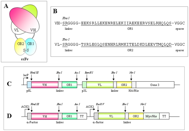
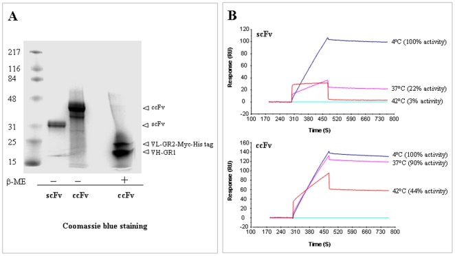
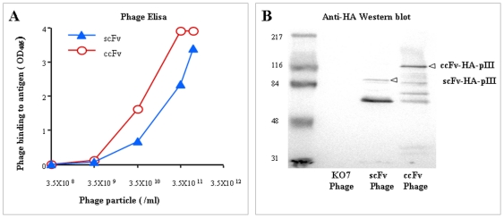
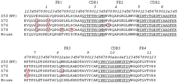
A Fab-like antibody binding unit, ccFv, in which a pair of heterodimeric coiled-coil domains was fused to V(H) and V(L) for Fv stabilization, was constructed for an anti-VEGF antibody. The anti-VEGF ccFv showed the same binding affinity as scFv but significantly improved stability and phage display level. Furthermore, phage display libraries in the ccFv format were constructed for humanization and affinity maturation of the anti-VEGF antibody. A panel of V(H) frameworks and V(H)-CDR3 variants, with a significant improvement in affinity and expressibility in both E. coli and yeast systems, was isolated from the ccFv phage libraries. These results demonstrate the potential application of the ccFv antibody format in antibody engineering.


Conflict of interest statement

Competing Interests: All authors are employees of Merck Co. & Inc., which acquired their previous employer, Abmaxis Inc., in 2006. There are no products in development or marketed products to declare. There are three US patents issued: US patents 6833441 (December 21, 2004), 7429652 (Sept 30, 2008) US patent 7667004 (February 24, 2010). This does not alter the authors' adherence to all the PLoS ONE policies on sharing data and materials. This manuscript has been reviewed and released by the Office of Scientific & Technical Information Clearance (OSTIC) of Merck for publication.

Figures

Figure 1
Figure 1. An overview of the coiled-coil Fv (ccFv) construct.
(A) The ccFv molecule design. The VH and VL are heterodimerized through the interaction of GR1 and GR2 coiled-coil domains. (B) The amino acid sequences of GR1, GR2, linker, and spacer. (C) pABMD5, the ccFv phage display vector. (D) pABMX168, the ccFv expression vector in Pichia.
Figure 2
Figure 2. The expression of the ccFv fragment in Pichia and the study of its stability.
(A) SDS-PAGE analysis of purified ccFv and scFv, under reducing (β-ME) and non-reducing conditions. ccFv showed an electrophoretic mobility of approximately 35 kDa. Under reducing conditions, two ccFv subunits corresponding to VL-GR2-Myc-His tag and VH-GR1 were observed. (B) A thermal stability comparison between ccFv and the corresponding scFv. The anti-VEGF ccFv and scFv fragments were incubated at 4°C (referred to as 100% binding activity), 37°C, and 42°C. Their remaining binding activity to the VEGF antigen was measured by Biacore at 25°C.
Figure 3
Figure 3. The functional phage display of the ccFv fragment.
(A) Phage ELISA was conducted using the phages displaying either the ccFv or scFv fragment encoding the same anti-VEGF antibody. The results demonstrated that ccFv was functionally assembled, expressed, and displayed on phages. The display level was higher by almost one order of magnitude than scFv. (B) A western blot of phages displaying ccFv and scFv fragments. The same amounts of phage particles displaying either the ccFv or scFv fragment were analyzed by SDS-PAGE and anti-HA western blotting. More intact ccFv-pIII fusion proteins were detected than the intact scFv-pIII fusion proteins, suggesting a higher ccFv phage display level.
Figure 4
Figure 4. The framework amino acid sequences of the leads selected from the ccFv humanization library for the anti-VEGF antibody.
All three leads, X72, X76, and X78, differed from WT at the E6Q and L11V mutations in the VH framework 1. The Kabat numbering scheme was used for residue numbering.
Figure 5
Figure 5. Improved expression of the leads identified from the ccFv humanization and maturation libraries for an anti-VEGF antibody.
The leads identified from the ccFv libraries were converted to scFv and expressed in Pichia. (A) SDS-PAGE analysis of the expression of the humanization leads (X72, X76, and X78) and the WT control. All three leads showed an expression level significantly higher than that of WT X50. (B) SDS-PAGE analysis of the expression of X96 and X97, which carried the combined mutations identified from both the humanization and the maturation libraries.

Similar articles

Cited by

References

    1. Kontermann RE. Alternative antibody formats. Curr Opin Mol Ther . 2010;12:176–183. - PubMed
    1. Nelson AL. Antibody fragments: hope and hype. MAbs . 2010;2:77. - PMC - PubMed
    1. Bird RE, Hardman KD, Jacobson JW, Johnson S, Kaufman BM, et al. Single-chain antigen-binding proteins. Science. 1988;242:423–426. - PubMed
    1. Huston JS, Levinson D, Mudgett-Hunter M, Tai MS, Novotny J, et al. Protein engineering of antibody binding sites: recovery of specific activity in an anti-digoxin single-chain Fv analogue produced in Escherichia coli. Proc Natl Acad Sci USA . 1998;85:5879–3883. - PMC - PubMed
    1. Kortt AA, Malby RL, Caldwell JB, Gruen LC, Ivancic N, et al. Recombinant anti-sialidase single-chain variable fragment antibody. Eur J Biochem . 1994;221:151–157. Characterization, formation ofdimer and higher-molecular-mass multimers and the solution of the crystal structure of the single-chain variable fragment/sialidase complex. - PubMed

Substances