. 2011 Apr 28;6(4):e19291.
doi: 10.1371/journal.pone.0019291.

αA-crystallin peptide SDRDKFVIFLDVKHF accumulating in aging lens impairs the function of α-crystallin and induces lens protein aggregation

Affiliations

αA-crystallin peptide SDRDKFVIFLDVKHF accumulating in aging lens impairs the function of α-crystallin and induces lens protein aggregation

Puttur Santhoshkumar et al. PLoS One. .

Abstract

Background: The eye lens is composed of fiber cells that are filled with α-, β- and γ-crystallins. The primary function of crystallins is to maintain the clarity of the lens through ordered interactions as well as through the chaperone-like function of α-crystallin. With aging, the chaperone function of α-crystallin decreases, with the concomitant accumulation of water-insoluble, light-scattering oligomers and crystallin-derived peptides. The role of crystallin-derived peptides in age-related lens protein aggregation and insolubilization is not understood.

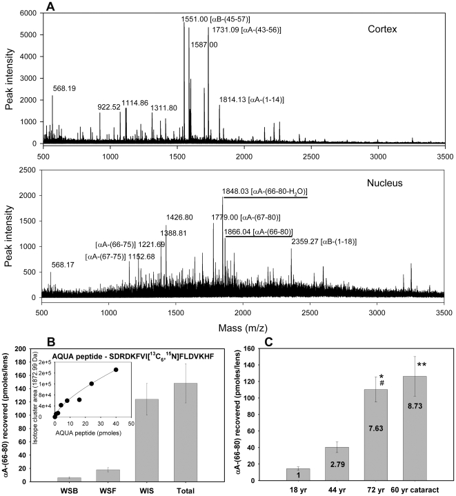
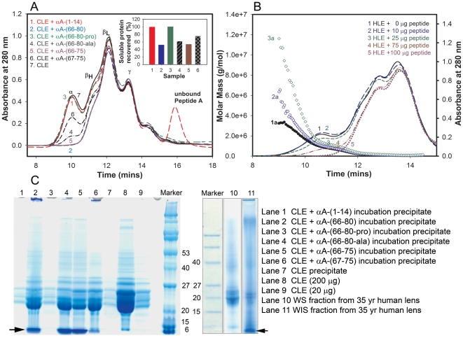
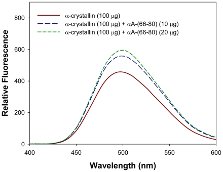
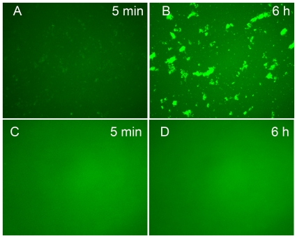
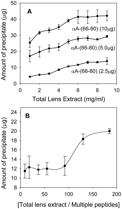
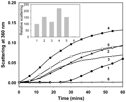
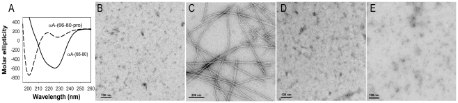
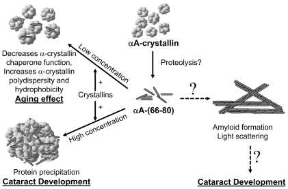
Methodology/principal findings: We found that αA-crystallin-derived peptide, (66)SDRDKFVIFLDVKHF(80), which accumulates in the aging lens, can inhibit the chaperone activity of α-crystallin and cause aggregation and precipitation of lens crystallins. Age-related change in the concentration of αA-(66-80) peptide was estimated by mass spectrometry. The interaction of the peptide with native crystallin was studied by multi-angle light scattering and fluorescence methods. High molar ratios of peptide-to-crystallin were favourable for aggregation and precipitation. Time-lapse recordings showed that, in the presence of αA-(66-80) peptide, α-crystallin aggregates and functions as a nucleus for protein aggregation, attracting aggregation of additional α-, β- and γ-crystallins. Additionally, the αA-(66-80) peptide shares the principal properties of amyloid peptides, such as β-sheet structure and fibril formation.

Conclusions/significance: These results suggest that crystallin-derived peptides such as αA-(66-80), generated in vivo, can induce age-related lens changes by disrupting the structure and organization of crystallins, leading to their insolubilization. The accumulation of such peptides in aging lenses may explain a novel mechanism for age-related crystallin aggregation and cataractogenesis.


Conflict of interest statement

Competing Interests: The authors have declared that no competing interests exist.

Figures

Figure 1
Figure 1. Distribution of the αA-(66-80) peptide in the lens.
(A) MALDI qTOF MS/MS analysis of LMW peptides isolated from the cortex and nucleus of a >70-year-old human lens as described under methods. The αA-(66-80) peptide characterized in this study is highlighted in the panel labeled – nucleus. (B) αA-(66-80) peptide concentrations in a >70-year-old lens fractions (WSB, water-soluble bound; WSF, water-soluble free; WIS, water-insoluble sonicated fractions). The amount of the peptide was estimated using 13C 15N-labelled spiked synthetic peptide standards (AQUA peptide) during peptide isolation. The inset figure is the standard curve for the isotope cluster area of the AQUA peptide used to determine in vivo peptide concentration. (C) Concentrations of αA-(66-80) peptide in human lenses of 18-, 44-, 60- and 72-year age groups. Lenses that were 60 years old were cataract lenses. Values within the bar show the multiple increased concentrations as compared to the 18-year-old lens. The values are mean ± SD of three analyses. * P<0.001 compared to 18 year, # P<0.01 compared to 44 year, ** P<0.001 compared to 18 and 44 year. P value determined using ANOVA. These results show an age-dependent increase in the concentration of αA-(66-80) peptide in human lenses and these peptides are associated with the WIS fraction. The cataract lenses show a higher level of αA-(66-80) peptide than the expected level of the peptide in age-matched non-cataract lenses.
Figure 2
Figure 2. The αA-(66-80) peptide–induced aggregation and precipitation of crystallins.
(A) Size-exclusion column elution profiles of soluble crystallins from WS CLE (200 µg) following incubation with or without different synthetic peptides (100 µg) for 16 hrs at 37°C. The chromatography was performed using a TSK G5000PWXL column (7.8 mm×30 cm) and the elution was monitored using a 280 nm absorption detector. The inset in the figure shows the amount of soluble protein recovered after peptide treatment and centrifugation at 3000 rpm for 10 min. Samples 1-7 in the inset correspond to samples 1-7 analyzed by the size-exclusion column. The results show that the effect of αA-(66-80) peptide is sequence specific. The addition of αA-(66-80), αA-(66-75) and αA-(67-75) peptides, present in human lens WIS fractions, causes precipitation of crystallins when incubated with CLE. (B) Effect of the peptide on 35-year-old human lens crystallins. HLE (190 µg) was incubated with various amounts of synthetic αA-(66-80) peptide for 16 h at 37°C. The samples were centrifuged, and the supernatants were chromatographed on a TSK G5000PWXL column connected to a multi-angle light scattering instrument (Wyatt Technology). Lines 1a, 2a and 3a show the distribution of molecular mass across the UV profile of samples 1, 2 and 3, respectively. Mass profiles of samples 4 and 5 could not be analyzed due to instrument limitations. The results show that the presence of increased amounts of αA-(66-80) peptide in the incubation mixture leads to increased aggregation, light scattering and precipitation. (C) SDS-PAGE analysis of precipitates of CLE and αA-derived peptides or substituted αA-peptides from panel A and the WS and WIS fractions from a 35-year-old human lens. The left arrow on lane 1 points to the precipitation of test peptides with crystallins and the right arrow on lane 11 points to the presence of peptides in human WIS fraction. The results of SDS-PAGE analysis show the precipitate formed when αA-crystallin-derived peptides are incubated with CLE that contains both the lens crystallins and the peptide tested. The data also confirm the presence of LMW protein fragments in the WIS fraction from human lenses.
Figure 3
Figure 3. Bis-ANS binding to α-crystallin treated with or without αA-(66-80).
α-Crystallin (100 µg) purified from a bovine lens extract was incubated with and without 10 or 20 µg of αA-(66-80) peptide in 50 mM phosphate buffer, pH 7.2 for 60 min and 10 µL of 20 mM bis-ANS prepared in 5% ethanol was added. Fluorescence was recorded after excitation at 390 nm in a Jasco FP750 spectrofluorometer. The fluorescence for αA-(66-80) was subtracted from the spectra for the complexes. These data show that the interaction of this peptide with α-crystallin leads to an overall increase in the hydrophobicity of the complex.
Figure 4
Figure 4. αA-(66-80) Peptide–induced HMW aggregate formation.
Aggregates formed after incubation mixtures of 25 µg αA-(66-80) peptide with 200 µg α-crystallin, as described under methods, were re-suspended with Alexa-488 labelled αB-crystallin (αBT162C-488) in 50 mM phosphate buffer, pH 7.2, and the mixture was incubated at 37°C. The protein sample was removed after 5 min and 6 hrs and placed on a pre-cleaned glass slide and observed under the Leica fluorescence microscope using blue filter. The image was captured at 20× magnification. (A) Sample after 5 min; (B) sample after 6 h. As a control, a mixture of αA-(66-80-Pro) peptide–treated α-crystallin was incubated overnight and an aliquot was mixed with αBT162C-488 and observed under fluorescence microscope at 5 min (C) and 6 h D). The results show that α-crystallin aggregates formed after incubation with αA-(66-80) peptide bind αB-crystallin. The findings suggest that peptide- α-crystallin acts as a nucleus for binding of αB-crystallin.
Figure 5
Figure 5. Effect of protein concentration on peptide-induced aggregation of human lens proteins.
(A) Lens crystallin fraction (0.5–9 mg) was incubated with 2.5–10 µg of αA-(66-80) peptide in 1 ml buffer at 37°C. The amount of protein precipitated after 24 h was estimated using Bio-Rad protein assay reagent (n = 3). The amount of precipitate formed increased when the reaction mixture contained increased amounts of peptide or lens extract. (B) Effect of a mixture of peptides on lens proteins. Different concentrations of dialyzed human lens extract were mixed with a mixture of peptides consisting of αA-(66-80), αA-(66-75), αA-(67-75), αB-(1-18) and βA3/A1-(102-117), 1.0 µg each, to obtain 5- to 200-fold excess (by weight) of crystallins. The mixtures were incubated at 37°C for 24 h in 1 ml of 50 mM phosphate buffer, pH 7.2. The precipitate formed was collected by centrifugation and estimated as above. The results show that the presence of several peptides in low concentration is sufficient to induce lens crystallin precipitation when the crystallin concentration is high. Therefore, an in vivo lens protein concentration of 400 mg/ml is likely to aggregate and precipitate even when the peptide concentration is 100's of times lower than that of crystallins.
Figure 6
Figure 6. The αA-(66-80) peptide functions as an anti-chaperone.
The effect of αA-(66-80) peptide on the chaperone activity of αB-crystallin against denaturing ADH (150 µg) was examined using the assay procedure described earlier . (1) ADH; (2) ADH+αA-(66-80) (25 µg); (3) ADH+αB (30 µg)+αA-(66-80) (25 µg); (4) ADH+αA-(66-80) (50 µg); (5) ADH+αB (30 µg)+αA-(66-80) (50 µg); and (6) ADH+αB (30 µg). The relative scattering by the samples at the 40-min time point is shown in the inset graph. As expected, ADH aggregation is suppressed in the presence of αA-crystallin. But ADH aggregation is increased in presence of αA-(66-80) peptide, suggesting that the peptide facilitates the aggregation of denaturing ADH. Additionally, the chaperone activity of αB-crystallin is diminished in reactions containing αA-(66-80) peptide, suggesting that the peptide interferes in the chaperone activity of αB-crystallin that exhibits anti-chaperone activity.
Figure 7
Figure 7. Amyloid-like characteristics of the αA-(66-80) peptide.
(A) Far-UV CD spectra of 0.1 mg/ml αA-(66-80) peptide and the Pro-substituted peptide in phosphate buffer, pH 7.2, obtained as described under methods. Proline substitution abolishes the β-sheet structure of the peptide. (B, C, D) TEM micrographs of peptides incubated in 50 mM phosphate buffer (pH 7.2) at 37°C. (B) αA-(66-80) at 0 min; (C) αA-(66-80) after 24 h; and (D) TEM of the Pro-substituted αA-(66-80) peptide after 24 h incubation. (E) TEM of the αA-(1-14) peptide after 24 h incubation. TEM studies show that αA-(66-80) peptide forms fibrils (D), whereas following Pro-substitution, the same peptide is unable to form fibrils (E), suggesting that a propensity to form β-sheet structure is necessary for fibril formation. The αA-(1-14) peptide did not form fibrils (E) and did not show β-sheet structure (data not shown).
Figure 8
Figure 8. Schematic of αA-(66-80) peptide–mediated lens aging and cataract development.
In vivo αA-(66-80) peptides interact with crystallins and affect the structure and organization of crystallins, leading to age-related changes and cataract development. At low concentrations, αA-(66-80) specifically interacts with α-crystallin, resulting in increased hydrophobicity and polydispersity and decreased chaperone activity of α-crystallin. At higher concentrations, αA-(66-80) binds to crystallins, leading to insolubilization of crystallins and cataract development. Localized accumulation of the αA-(66-80) peptide has the potential to form light-scattering amyloid-like structures. However, the latter is less likely because the interaction of the peptide with crystallins suppresses the formation of fibrils.

Similar articles

Cited by

References

    1. Bloemendal H, de Jong W, Jaenicke R, Lubsen NH, Slingsby C, et al. Ageing and vision: structure, stability and function of lens crystallins. Prog Biophys Mol Biol. 2004;86:407–485. - PubMed
    1. Horwitz J. Alpha-crystallin can function as a molecular chaperone. Proc Natl Acad Sci U S A. 1992;89:10449–10453. - PMC - PubMed
    1. Horwitz J, Emmons T, Takemoto L. The ability of lens alpha crystallin to protect against heat-induced aggregation is age-dependent. Curr Eye Res. 1992;11:817–822. - PubMed
    1. Bron AJ, Vrensen GF, Koretz J, Maraini G, Harding JJ. The ageing lens. Ophthalmologica. 2000;214:86–104. - PubMed
    1. Sharma KK, Santhoshkumar P. Lens aging: effects of crystallins. Biochim Biophys Acta. 2009;1790:1095–1108. - PMC - PubMed

Publication types