. 2011 Apr 28;6(4):e19411.
doi: 10.1371/journal.pone.0019411.

Structural basis for variant-specific neuroligin-binding by α-neurexin

Affiliations

Structural basis for variant-specific neuroligin-binding by α-neurexin

Hiroki Tanaka et al. PLoS One. .

Abstract

Neurexins (Nrxs) are presynaptic membrane proteins with a single membrane-spanning domain that mediate asymmetric trans-synaptic cell adhesion by binding to their postsynaptic receptor neuroligins. α-Nrx has a large extracellular region comprised of multiple copies of laminin, neurexin, sex-hormone-binding globulin (LNS) domains and epidermal growth factor (EGF) modules, while that of β-Nrx has but a single LNS domain. It has long been known that the larger α-Nrx and the shorter β-Nrx show distinct binding behaviors toward different isoforms/variants of neuroligins, although the underlying mechanism has yet to be elucidated. Here, we describe the crystal structure of a fragment corresponding to the C-terminal one-third of the Nrx1α ectodomain, consisting of LNS5-EGF3-LNS6. The 2.3 Å-resolution structure revealed the presence of a domain configuration that was rigidified by inter-domain contacts, as opposed to the more common flexible "beads-on-a-string" arrangement. Although the neuroligin-binding site on the LNS6 domain was completely exposed, the location of the α-Nrx specific LNS5-EGF3 segment proved incompatible with the loop segment inserted in the B+ neuroligin variant, which explains the variant-specific neuroligin recognition capability observed in α-Nrx. This, combined with a low-resolution molecular envelope obtained by a single particle reconstruction performed on negatively stained full-length Nrx1α sample, allowed us to derive a structural model of the α-Nrx ectodomain. This model will help us understand not only how the large α-Nrx ectodomain is accommodated in the synaptic cleft, but also how the trans-synaptic adhesion mediated by α- and β-Nrxs could differentially affect synaptic structure and function.


Conflict of interest statement

Competing Interests: The authors have declared that no competing interests exist.

Figures

Figure 1
Figure 1. Structural and functional overlap between α- and β-Neurexin.
(A) Domain architecture. The ectodomain of α-Nrx can be divided into three repeating units (I∼III), each containing an EGF module flanked by two LNS domains. Module III corresponds to the fragment crystallized in the current study. The LNS6 domain is followed by a linker segment of 102 residues, a transmembrane domain (TM), and a cytoplasmic tail containing a PDZ-binding domain at the C-terminus. Locations of the five alternative splicing sites are indicated by arrows. β-Nrx shares the identical sequence as α-Nrx after LNS6, but contains a unique segment of 37 residues at the N-terminal preceded by the signal peptide (SP). (B) NX1α(III) and NX1αEC exhibit identical binding selectivity toward the NL1 splice variants. Binding between the Myc-tagged fragments of Nrx1α and the hGH-fusion NL1 variants were evaluated using a solid-phase binding assay followed by detection with an anti-Myc antibody. Note that all of the Nrx fragments bound well to NL1_B−, while only the NX1β bound to NL1_B+. A major immunoreactive band of 180 kDa (indicated by an asterisk) corresponds to mouse IgG dissociated from antibody beads.
Figure 2
Figure 2. Structure of NX1α(III).
(A) Two different views of the NX1α(III) structure in ribbon presentation, with the LNS5 domain colored in green, the EGF domain in magenta, and the LNS6 domain in cyan. A Ca2+ and an N-acetylglucosamine (GlcNac) residue attached to Asn 1186 are shown as a yellow sphere and a stick model, respectively. (B) Molecular surface of NX1α(III) colored and viewed as in (A). Residues that constitute the NL1-binding site in Nrx1β are colored in red. (C) Multiple-sequence alignment of LNS and EGF domains of Nrx1α. Residue numbers are based on the shortest versions (i.e., without any splice site insertions) of the bovine Nrx1α sequence. For those domains that have structural information, secondary structural elements are highlighted in cyan (β-strands) and red (α-helices), respectively. A segment corresponding to the inserted splicing site 1 (SS1), which was contained in the construct used in this study but which was excluded from the numbering, is highlighted in magenta. Trp1065 in EGF3, which plays an important role in the interdomain interaction, is highlighted in yellow. Conserved disulfide bonds are indicated by gray lines.
Figure 3
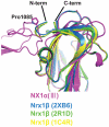
Figure 3. Difference in the N-terminal trajectory of the LNS6 domain between Nrx1α and Nrx1β.
Previously determined structures of Nrx1β (PDB ID: 2R1D(green), 1C4R(yellow), 2XB6(blue)), and the LNS6 domain of NX1α(III) (magenta) are superposed. The chain-diverting Pro1085 in NX1α(III) is shown as a stick model.
Figure 4
Figure 4. The structures of the LNS5 and LNS6 domains.
(A) Close-up views of the Ca2+-binding pockets of LNS5 (left) and LNS6 (right). In the LNS6 domain, a Ca2+ (yellow sphere) is coordinated by four protein ligands and two water molecules. The same site in LNS5 is occupied by a water molecule. (B) Inter-domain contacts at the LNS5-EGF3 (left) and EGF3-LNS6 (right) junctions. LNS domains are shown in surface rendering while the EGF domain emerging from them is shown as a ribbon model, all colored as in Fig. 2A. Side-chains of the residues directly participating in the interactions are represented as a stick model, and hydrogen bonds are depicted as dotted lines.
Figure 5
Figure 5. NX1α(III) uses the same NL1-binding interface as NX1β.
The binding of the MycHis-tagged NX1α(III) with the indicated mutations to the hGH_NL1_B− was evaluated by a solid-phase binding assay as in Figure 1B. NX1α(III) proteins bound by NL1, through the anti-hGH beads, were visualized with a Western blot using an anti-Myc antibody (top). The same culture supernatants were precipitated with Ni-NTA agarose to confirm the similar expression levels of each mutant (bottom). The migration position for the tagged NX1α(III) is indicated with arrowheads.
Figure 6
Figure 6. Structural model of the NX1α(III)/NL1 complex.
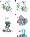
(A) Simple superposition of the NX1α(III) onto the Nrx1β/NL1 heterotetramer structure (PDB ID : 3B3Q) at the LNS6 domain, resulting in a steric clash between the LNS5 domain and the NL1 (red dotted circle). In the NX1α(III) structure, the putative pivot point (Pro1085) and the domain-locking Trp1065 are shown as stick models. (B) Hypothetical model of the NX1α(III)/NL1 complex after simulated domain rotation. The LNS5+EGF3 segment in NX1α(III) was rotated 18° clockwise around the Pro1085, relieving the clash. (C) The binding interface of the Nrx1β/NL1 complex demonstrates considerable plasticity. Four pairs of Nrx1β/NL1 heterodimeric complexes were excised from the reported 2∶2 complex structures (PDB ID: 3B3Q and 3BIW), and were superposed at the NL1 molecule. (D) Potential effect of splice site B insertion in NL1 on Nrx binding. Two different views of the same structural model as in (A) are shown, with the Nrx-binding NL1 residues painted in blue. For clarity, the LNS5 domain is omitted. The insertion point for the 9-residue splicing site B sequence (between Glu297-Gly298 in NL1, painted in yellow) is adjacent to the interface. In the side view (lower panel), a dotted line represents the estimated route of the inserted loop containing the bulky N-linked sugar chain.
Figure 7
Figure 7. Electron microscopic analysis of the Nrx1α ectodomain (NX1αEC).
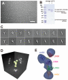
(A) A raw image of the negatively stained NX1αEC. Scale bar: 50 nm. (B) Spontaneous degradation of purified NX1αEC. SDS-PAGE analysis of NX1αEC protein immediately after purification (left) and again after 2 months storage at 4°C (right) shows that the protein undergoes proteolytic cleavage to produce N-terminal (30 kDa) and C-terminal (110-kDa) fragments. (C) Two-dimensional class averages (upper row images) obtained from multiple electron micrographs and corresponding projection views produced from the 3-D volume map (lower row images). Scale bar, 10 nm. (D) Three-dimensional reconstruction of the NX1αEC created from multiple oriented particles. (E) Predicted 3D domain organization within the NX1αEC fragment. Atomic coordinates for NX1α(III) are manually fitted to the densities corresponding to the LNS5-6 and LNS3-4 segments, keeping the C-terminus of LNS4 and the N-terminus of LNS5 close enough to be connected. Domain connectivity was also considered when fitting the LNS2 domain structure (PDB ID: 2H0B) into the density at the bottom. LNS1+EGF1 segment disappeared in the reconstructed volume was not assigned.
Figure 8
Figure 8. Schematic rendering of the Nrx1α/NL1 complex in the synaptic cleft.
Hypothetical NL1 dimer simultaneously bound by Nrx1β and Nrx1α from opposite sides are depicted in two different views. For Nrx1α-NL1 association, structural models of the NX1α(III)/NL1 complex (Fig. 6B) and the NX1αEC (Fig. 7E) are combined. In this rendering, LNS1+EGF1 segment of the Nrx1α was modeled but the position was determined arbitrary. The linker segments connecting toward cell membrane in both proteins are represented by dotted lines. The Nrx1/NL1 complex structure is drawn roughly in scale to the length of the synaptic cleft (∼200 Å).

Similar articles

Cited by

References

    1. Craig AM, Kang Y. Neurexin-neuroligin signaling in synapse development. Curr Opin Neurobiol. 2007;17:43–52. - PMC - PubMed
    1. Dean C, Dresbach T. Neuroligins and neurexins: linking cell adhesion, synapse formation and cognitive function. Trends Neurosci. 2006;29:21–29. - PubMed
    1. Dean C, Scholl FG, Choih J, DeMaria S, Berger J, et al. Neurexin mediates the assembly of presynaptic terminals. Nat Neurosci. 2003;6:708–716. - PMC - PubMed
    1. Graf ER, Zhang X, Jin SX, Linhoff MW, Craig AM. Neurexins induce differentiation of GABA and glutamate postsynaptic specializations via neuroligins. Cell. 2004;119:1013–1026. - PMC - PubMed
    1. Nam CI, Chen L. Postsynaptic assembly induced by neurexin-neuroligin interaction and neurotransmitter. Proc Natl Acad Sci U S A. 2005;102:6137–6142. - PMC - PubMed

Publication types

Associated data

LinkOut - more resources