

Review
doi: 10.1590/s1807-59322011000300023.
Atypical mole syndrome and dysplastic nevi: identification of populations at risk for developing melanoma - review article
Affiliations
- PMID: 21552679
- PMCID: PMC3072014
- DOI: 10.1590/s1807-59322011000300023
Item in Clipboard
Review
Atypical mole syndrome and dysplastic nevi: identification of populations at risk for developing melanoma - review article
Clinics (Sao Paulo).
2011.
Abstract
Atypical Mole Syndrome is the most important phenotypic risk factor for developing cutaneous melanoma, a malignancy that accounts for about 80% of deaths from skin cancer. Because the diagnosis of melanoma at an early stage is of great prognostic relevance, the identification of Atypical Mole Syndrome carriers is essential, as well as the creation of recommended preventative measures that must be taken by these patients.
Figures
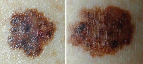
Macroscopic image of two melanocytic lesions whose characteristics are superimposed by the ABCD rule (asymmetry, irregular borders, varied coloration, diameter greater than 6 mm): Left - dysplastic nevus; Right – cutaneous melanoma.
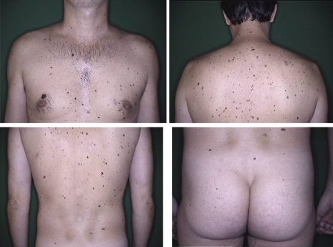
Body mapping of a male patient, 34 years of age, 256 nevi selected for follow-up. Presents 3 criteria for AMS: more than 100 nevi, presence of more than 2 clinically dysplastic nevi and nevi on the buttocks.
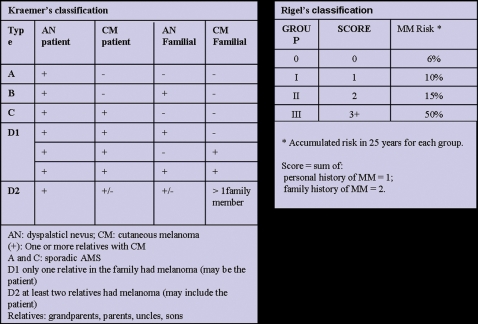
Kraemer and Rigel Classifications.
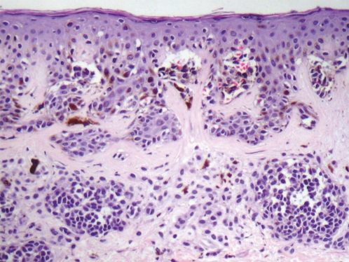
Histopathologic exam of atypical nevus showing focal atypia of melanocytes, fusion of epithelial cones and concentric lamellar fibrosis. Optical microscopy image with high magnification (40X).
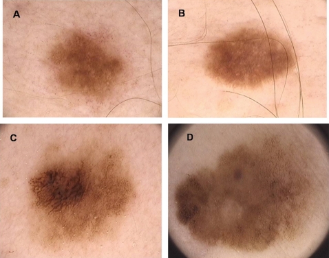
Digital dermoscopy (20X) of melanocytic lesions that have similar dermatoscopic appearance (A ∼ B; C ∼ D): A) compound melanocytic nevus; B) dysplastic nevus; C) dysplastic junctional nevus with severe atypia, D) thin melanoma.
Similar articles
-
The dangers of atypical mole (dysplastic nevus) syndrome. Teaching at-risk patients to protect themselves from melanoma.Postgrad Med. 1999 Jun;105(7):147-8, 151-2, 154 passim. doi: 10.3810/pgm.1999.06.624. Postgrad Med. 1999. PMID: 10376056
-
Atypical mole syndrome: risk factor for cutaneous malignant melanoma and implications for management.J Am Acad Dermatol. 1995 Mar;32(3):479-94. doi: 10.1016/0190-9622(95)90073-x. J Am Acad Dermatol. 1995. PMID: 7868720 Review.
-
[Dysplastic nevus].Presse Med. 1990 Feb 17;19(6):239-41. Presse Med. 1990. PMID: 2138293 French. No abstract available.
-
Dysplastic nevus (atypical nevus).An Bras Dermatol. 2010 Nov-Dec;85(6):863-71. doi: 10.1590/s0365-05962010000600013. An Bras Dermatol. 2010. PMID: 21308311 Review. English, Portuguese.
-
Dysplastic nevi. Markers for increased risk for melanoma.Cancer. 1989 Jan 15;63(2):386-9. doi: 10.1002/1097-0142(19890115)63:2<386::aid-cncr2820630231>3.0.co;2-6. Cancer. 1989. PMID: 2910446
Cited by
-
Recognizing Histopathological Simulators of Melanoma to Avoid Misdiagnosis.Cureus. 2022 Jun 20;14(6):e26127. doi: 10.7759/cureus.26127. eCollection 2022 Jun. Cureus. 2022. PMID: 35875272 Free PMC article. Review.
-
Multiple primary cutaneous melanomas in patients with FAMMM syndrome and sporadic atypical mole syndrome (AMS): what's worse?Wien Med Wochenschr. 2014 Aug;164(15-16):302-7. doi: 10.1007/s10354-014-0295-8. Epub 2014 Aug 6. Wien Med Wochenschr. 2014. PMID: 25096163
-
AC-1001 H3 CDR peptide induces apoptosis and signs of autophagy in vitro and exhibits antimetastatic activity in a syngeneic melanoma model.FEBS Open Bio. 2016 Jul 15;6(9):885-901. doi: 10.1002/2211-5463.12080. eCollection 2016 Sep. FEBS Open Bio. 2016. PMID: 27642552 Free PMC article.
-
Overview of familial syndromes with increased skin malignancies.Arch Dermatol Res. 2023 May;315(4):707-727. doi: 10.1007/s00403-022-02447-8. Epub 2022 Nov 7. Arch Dermatol Res. 2023. PMID: 36342513 Review.
-
Nevus Variations in the Jordanian Population: Effects of Age, Medical Conditions, Environment, Congenital, Inherited, and Genetic Factors.Clin Cosmet Investig Dermatol. 2024 Jan 4;17:17-29. doi: 10.2147/CCID.S433447. eCollection 2024. Clin Cosmet Investig Dermatol. 2024. PMID: 38193026 Free PMC article.
References
-
- Newton JA, Bataille V, Griffiths K, Squire JM, Sasieni P, Cuzick J, et al. How common is the atypical mole syndrome phenotype in apparently sporadic melanoma. J Am Acad Dermatol. 1993;29:989–96. 10.1016/0190-9622(93)70279-3 - DOI - PubMed
-
- Gandini S, Sera F, Cattaruzza MS, Pasquini P, Abeni D, Boyle P, et al. Meta-analysis of risk factors for cutaneous melanoma: I. Common and atypical naevi. Eur J Cancer. 2005;41:28–44. - PubMed
-
- Slade J, Marghoob AA, Salopek TG, Rigel DS, Kopf AW, Bart RS. Atypical mole syndrome: Risk factor for cutaneous malignant melanoma and implications for management. J Am Acad Dermatol. 1995;32:479–94. 10.1016/0190-9622(95)90073-X - DOI - PubMed
-
- Miller AJ, Mihm MC., Jr Melanoma. N Engl J Med. 2006;355:51–65. - PubMed
-
- Thompson JF, Scolyer RA, Kefford RF. Cutaneous Melanoma. Lancet. 2005;365:687–701. - PubMed
Publication types
MeSH terms
LinkOut - more resources
Full Text Sources
Medical
