. 2011 May;118(5):795-808.
doi: 10.1007/s00702-011-0653-2. Epub 2011 May 7.

Parkinson's disease-linked LRRK2 is expressed in circulating and tissue immune cells and upregulated following recognition of microbial structures

Affiliations

Parkinson's disease-linked LRRK2 is expressed in circulating and tissue immune cells and upregulated following recognition of microbial structures

Mansoureh Hakimi et al. J Neural Transm (Vienna). 2011 May.

Abstract

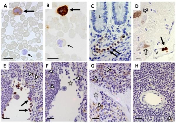
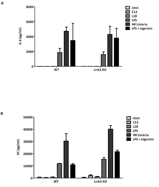
Sequence variants at or near the leucine-rich repeat kinase 2 (LRRK2) locus have been associated with susceptibility to three human conditions: Parkinson's disease (PD), Crohn's disease and leprosy. As all three disorders represent complex diseases with evidence of inflammation, we hypothesized a role for LRRK2 in immune cell functions. Here, we report that full-length Lrrk2 is a relatively common constituent of human peripheral blood mononuclear cells (PBMC) including affinity isolated, CD14(+) monocytes, CD19(+) B cells, and CD4(+) as well as CD8(+) T cells. Up to 26% of PBMC from healthy donors and up to 43% of CD14(+) monocytes were stained by anti-Lrrk2 antibodies using cell sorting. PBMC lysates contained full-length (>260 kDa) and higher molecular weight Lrrk2 species. The expression of LRRK2 in circulating leukocytes was confirmed by microscopy of human blood smears and in sections from normal midbrain and distal ileum. Lrrk2 reactivity was also detected in mesenteric lymph nodes and spleen (including in dendritic cells), but was absent in splenic mononuclear cells from lrrk2-null mice, as expected. In cultured bone marrow-derived macrophages from mice we made three observations: (i) a predominance of higher molecular weight lrrk2; (ii) the reduction of autophagy marker LC3-II in (R1441C)lrrk2-mutant cells (<31%); and (iii) a significant up-regulation of lrrk2 mRNA (>fourfold) and protein after exposure to several microbial structures including bacterial lipopolysaccharide and lentiviral particles. We conclude that Lrrk2 is a constituent of many cell types in the immune system. Following the recognition of microbial structures, stimulated macrophages respond with altered lrrk2 gene expression. In the same cells, lrrk2 appears to co-regulate autophagy. A pattern recognition receptor-type function for LRRK2 could explain its locus' association with Crohn's disease and leprosy risk. We speculate that the role of Lrrk2 in immune cells may also be relevant to the susceptibility of developing PD or its progression.


Figures

Figure 1
Figure 1. Lrrk2 reactivity in mammalian leukocytes using immunocytochemistry and immunohistochemistry
(A-D) Lrrk2 reactivity was detected in select leukocytes of peripheral blood smears (A-B; healthy controls; n=5), and in intravascular leukocytes of the submucosa in the distal ileum (C; neurological controls; n=3) and human midbrain (D) from a Parkinson disease donor, as denoted by large arrows. Small arrows point to unstained and poorly stained leukocytes; open arrows identify autofluorescent neuromelanin in dopaminergic cells of the midbrain. V, indicates vessel; L, lumen of the gut. Bar length equals 12 μm; original magnification, 60x. Lrrk2 signal was detected by affinity-purified, polyclonal anti-Lrrk2 antibody [“HL-2” raised against a synthetic peptide comprising residues 2508-2527 of the human, wild-type Lrrk2 (Berger et al. 2010)]. (E-H) The specificity of the Lrrk2 antibody used in A-D was confirmed in spleen sections from age-matched, wild-type (E-F) and lrrk2 knock-out (G-H) mice (n=4 each). Parallel sections were developed with anti-Lrrk2 HL-2 as primary antibody (E; G) or without primary antibody (F; H). Arrows depict lrrk2-immunoreactive leukocytes in the spleen; open arrow heads show autofluorescent cytoplasmic granules seen irrespective of antibody use; open asterisks denote groups of unstained leukocytes (E-H). Bar length equals 20 μm; original magnification, 40x.
Figure 2
Figure 2. Lrrk2 reactivity in human peripheral blood mononuclear cells and isolated monocytes
(A-B) Lrrk2 reactivity was detected in peripheral blood mononuclear cells (PBMC) (A) and anti-CD14+ monocytes (B) that were isolated by Ficoll separation and magnetic beads (carrying anti-CD14 antibody), respectively, from healthy adult controls (n=3) (Vranjkovic et al. 2011). FACS sorting of fixed and permeabilized cells was carried out using monoclonal anti-Lrrk2 antibody MJFF-1(C5-8). In each panel, a selection of cells is made by the forward and side scatter light properties (horizontal and vertical axis respectively in the left diagram) and a histogram (left diagram, horizontal and vertical axis represent the number of the cells counted and fluorescence respectively) of the fluorescence (IgG) profile of the Lrrk2 is generated to show the degree of the antibody binding. Red vertical lines show mean fluoresce of unstained cells. PBMC and monocytes were collected from two different individuals. Note, only 0.58% of PBMC (A) and 0.81% of monocytes (B) were gated in the absence of anti-Lrrk2 antibody. However, following the optimization of primary and secondary antibody concentrations the percentage of Lrrk2-positive cells measured 19.04 (A2), 24.54 (A3), and 25.71 (A4) for PBMC; and 1.8 (B2), 10.31 (B3), and 43.17 (B4). Thus, staining with anti-Lrrk2 revealed that <25.71% of all PBMC (A) and <43.17% of all monocytes (B) were successfully gated.
Figure 3
Figure 3. Detection of full-length Lrrk2 protein in isolated monocytes, B-cells and T-cells
(A-F) Peripheral blood mononuclear cells (PBMC; A-B) were obtained by Ficoll separation from healthy adult controls (n=5) and separated from erythrocytes (RBC) and granulocytes. PBMC were further processed using antibodies to CD14 (monocyte marker; in C), CD4 and CD8 (both T-cell markers; in panels D-E), and to CD19 (B-cell marker; E), which were conjugated to and immobilized by magnetic beads (see Materials and Methods) (Vranjkovic et al. 2011). Isolated subpopulations of leukocytes were lysed and then subjected to denaturing SDS/PAGE under reducing conditions, followed by Western blotting. Transgenic flies expressing full-length hLRRK2 cDNA (tg) and their wild-type, non-transgenic littermates (wt) were used as positive and negative controls, respectively. EBV-transformed, B-cell derived lymphoblasts from healthy control and Parkinson disease donors (n=5) were run in parallel as additional controls (panel F). Immunoblot in A was probed with affinity-purified, polyclonal anti-human Lrrk2 antibody (HL-2) raised against residues 2508-2527; probing with a monoclonal rabbit anti-Lrrk2 antibody [MJFF-1] is shown in B. Note, polyclonal anti-Lrrk2 antibody NB300-268 was used for membranes C to F. Anti-acid β-glucosidase was used as an independent marker of peripheral monocytes (see bottom panel in C). All membranes were stripped and reprobed with anti-β-actin antibody as loading control. Note, the robust and specific detection of endogenous, full-length Lrrk2 proteins (245-260 kDa) and higher molecular weight (HMW) species thereof in human PBMC, monocytes, B-cells and T-cells. There, CD19+ B-cells showed a slightly stronger signal for Lrrk2 when compared to isolated CD4+ T-cells enriched from the same donor (E).
Figure 4
Figure 4. A likely role for Lrrk2 during autophagy and pathogen response in macrophages
(A-B) SDS/PAGE under denaturing, reducing conditions followed by immunoblotting of murine bone marrow-derived macrophages (BMDM) that were cultured for 48 hrs without stimulation. Lysate of human EBV-transformed lymphoblasts from a control donor was loaded as control. Lanes 2-4 show lysates of BMDM prepared in parallel from age-matched mice of three distinct lrrk2 genotypes: wild type (WT); heterozygous (HET); and homozygous knock-in (KI) mice expressing mutant R1441Clrrk2. Note, the robust expression of HMW lrrk2 in murine BMDM, as detected by monoclonal anti-Lrrk2 antibody [MJFF-4] in A. Sister aliquots of the same lysates were probed with an antibody to autophagy markers LC3-I and LC3-II (B). All membranes were stripped and reprobed with anti-β-actin antibody as loading control. Note, the reduction of LC3-II levels in mutant R1441Clrrk2-expressing BMDM compared to WT cells (≥31%; see text for details). (C) BMDM from WT mice were stimulated for 5 hrs with different Toll-like receptor (TLR) agonists: Pam3CSK4 (P3S) for TLR2; Poly I:C (PIC) for TLR3; LPS for TLR4; R837 for TLR7; and CpG for TLR9. Quantification of lrrk2 mRNA was performed using the ΔΔCT method (relative to 18S) and compared to levels in unstimulated BMDM cells. * denotes significant up-regulation (p<0.05 using ANOVA followed by Tukey test, n=3). For comparison, TNFα was upregulated in all samples >30-fold (not shown). (D) BMDM cultures from homozygous (R1441C / R1441C) and heterozygous (R1441C / WT) mice were stimulated with different inflammatory stimulants. Macrophages were transduced with a lentivirus expressing eGFP 48 hours prior to stimulations. Stimulants were added 24hrs after plating of cells. Note, transduction by virus caused an upregulation in lrrk2 protein levels when compared to the untransduced BMDM (compares lane 10 with lanes 2). EBV-lymph and Rapa refer to EBV-transformed lymphoblast and treatment with rapamycin, respectively. See Result section for further details.
Figure 5
Figure 5. Lrrk2 protein expression does not control IL-6 and KC cytokine signaling
(A-B) Screening of lrrk2 genotype-dependent cytokine signaling by cultured BMDM following exposure to different stimulants was explored by probing the release of interleukin-6 (IL-6; pg/ml in A-B) and keratinocyte-derived chemokine (KC; pg/ml; Suppl. Fig. 2) by ELISA. BMDM were isolated from lrrk2 knock-out (KO), and from age-matched, wild-type (WT) littermates (12 weeks-old; n=2 for each genotype) and cultured for 16 hrs (each supernatant analyzed in quadruplicates). Similarly, stimulants were added 24 hrs after plating. No specific lrrk2 expression-associated differences were detected in the conditioned media M for the production and release rate of KC and IL-6 (for additional genotypes, see also Suppl. Fig.2).

Similar articles

Cited by

References

    1. Alcaïs A, Mira M, Casanova JL, Schurr E, Abel L. Genetic dissection of immunity in leprosy. Curr Opin Immunol. 2005;17:44–48. - PubMed
    1. Alegre-Abarrategui J, Christian H, Lufino MM, Mutihac R, Venda LL, Ansorge O, Wade-Martins R. LRRK2 regulates autophagic activity and localizes to specific membrane microdomains in a novel human genomic reporter cellular model. Hum Mol Genet. 2009;18:4022–4034. - PMC - PubMed
    1. Aleyasin H, Rousseaux MW, Marcogliese PC, Hewitt SJ, Irrcher I, Joselin AP, Parsanejad M, Kim RH, Rizzu P, Callaghan SM, Slack RS, Mak TW, Park DS. DJ-1 protects the nigrostriatal axis from the neurotoxin MPTP by modulation of the AKT pathway. Proc Natl Acad Sci USA. 2010;107:3186–3191. - PMC - PubMed
    1. Barrett JC, Hansoul S, Nicolae DL, Cho JH, Duerr RH, Rioux JD, Brant SR, Silverberg MS, Taylor KD, Barmada MM, Bitton A, Dassopoulos T, Datta LW, Green T, Griffiths AM, Kistner EO, Murtha MT, Regueiro MD, Rotter JI, Schumm LP, Steinhart AH, Targan SR, Xavier RJ, NIDDK IBD Genetics Consortium. Libioulle C, Sandor C, Lathrop M, Belaiche J, Dewit O, Gut I, Heath S, Laukens D, Mni M, Rutgeerts P, Van Gossum A, Zelenika D, Franchimont D, Hugot JP, de Vos M, Vermeire S, Louis E, Belgian-French IBD Consortium. Wellcome Trust Case Control Consortium. Cardon LR, Anderson CA, Drummond H, Nimmo E, Ahmad T, Prescott NJ, Onnie CM, Fisher SA, Marchini J, Ghori J, Bumpstead S, Gwilliam R, Tremelling M, Deloukas P, Mansfield J, Jewell D, Satsangi J, Mathew CG, Parkes M, Georges M, Daly MJ. Genome-wide association defines more than 30 distinct susceptibility loci for Crohn’s disease. Nat Genet. 2008;40:955–962. - PMC - PubMed
    1. Berger Z, Smith KA, Lavoie MJ. Membrane localization of LRRK2 is associated with increased formation of the highly active LRRK2 dimer and changes in its phosphorylation. Biochemistry. 2010;49:5511–5523. - PMC - PubMed

Publication types

MeSH terms

Substances