. 2011 Jun 8;11(6):2560-6.
doi: 10.1021/nl201400z. Epub 2011 May 10.

Copper selenide nanocrystals for photothermal therapy

Affiliations

Copper selenide nanocrystals for photothermal therapy

Colin M Hessel et al. Nano Lett. .

Abstract

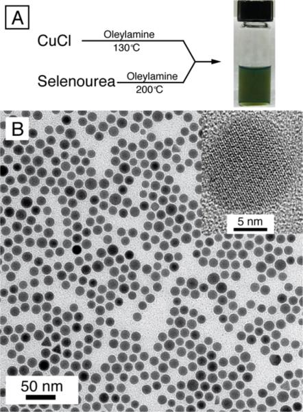
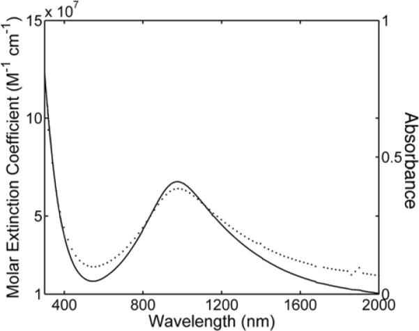
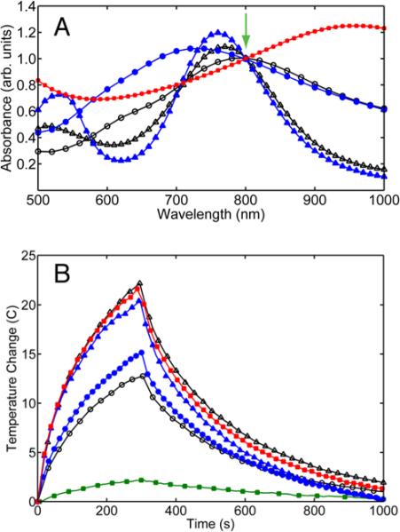
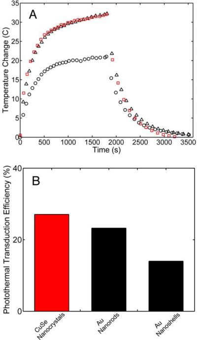
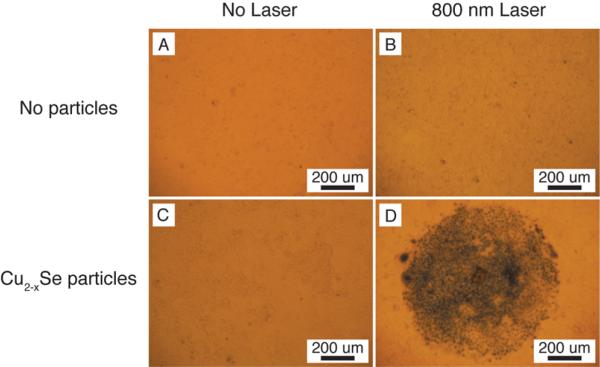
Ligand-stabilized copper selenide (Cu(2-x)Se) nanocrystals, approximately 16 nm in diameter, were synthesized by a colloidal hot injection method and coated with amphiphilic polymer. The nanocrystals readily disperse in water and exhibit strong near-infrared (NIR) optical absorption with a high molar extinction coefficient of 7.7 × 10(7) cm(-1) M(-1) at 980 nm. When excited with 800 nm light, the Cu(2-x)Se nanocrystals produce significant photothermal heating with a photothermal transduction efficiency of 22%, comparable to nanorods and nanoshells of gold (Au). In vitro photothermal heating of Cu(2-x)Se nanocrystals in the presence of human colorectal cancer cell (HCT-116) led to cell destruction after 5 min of laser irradiation at 33 W/cm(2), demonstrating the viabilitiy of Cu(2-x)Se nanocrystals for photothermal therapy applications.


Figures

Figure 1
Figure 1
(A) Reaction scheme and a photograph of oleylamine-capped Cu2−xSe nanocrystals dispersed in chloroform; (B) TEM images of copper selenide nanocrystals. The high resolution TEM image in the inset of (B) shows the crystalline Cu2−xSe core of the nanocrystals. The average diameter of the nanocrystals is 16±1 nm.
Figure 2
Figure 2
Amphiphilic polymer encapsulation of Cu2−xSe nanocrystals. Combining oleylamine passivated Cu2−xSe nanocrystals and the amphiphilic poly(maleic anhydride)-based polymer leads to encapsulation of the Cu2−xSe nanocrystals with a hydrophilic exterior. The distal carboxyl groups on the surface facilitate dispersibility in aqueous media.
Figure 3
Figure 3
Absorbance (dotted line) and molar extinction coefficient (solid line) for Cu2−xSe nanocrystals plotted against wavelength. The absorbance spectrum of polymer coated Cu2−xSe nanocrystals in water (37 mg/L) reaches a maximum at 970 nm. The molar extinction coefficient was calculated experimentally using Cu2−xSe solutions of chloroform, and is given per mole of 16 nm Cu2−xSe nanocrystals (see Supporting Information for calculations). The molar extinction coefficient reaches a maximum of 7.7×10⁷ M−1 cm−1 at 970 nm.
Figure 4
Figure 4
(A) Absorbance spectra of polymer-coated Cu2−xSe nanocrystals (red solid square), commercial Au nanoshells (blue solid circle) and Au nanorods (blue solid triangle), and synthesized Au nanoshells (black hollow circle) and Au nanorods (black hollow triangle) dispersed in deionized water. All solutions were normalized to an optical density equal to 1.0 at 800 nm (green arrow). (B) The photothermal response of the dispersions in (A) obtained by irradiating 300 μL aliquots of each solution for 5 min with an 800 nm diode laser (6 mm spot size, fluence of 2 W/cm2). The temperature was monitored with an infrared imaging camera. The laser heating of the water contributes approximately 2.5°C to the overall change in temperature in 5 min (green solid square).
Figure 5
Figure 5
TEM images of Au nanoshells and nanorods that were purchased from a commercial supplier and synthesized in-house. Synthesized Au nanoshells (A) are 135 nm in diameter, with an approximate shell thickness of 10 nm, while commercial Au nanoshells (B) are 145 nm in diameter, with a 7.5 nm thick Au shell. Synthesized nanorods (C) are 49 × 13 nm (aspect ratio: 3.8) and commercial nanorods (D) are 23 × 7 nm (aspect ratio: 3.3).
Figure 6
Figure 6
Steady state heating data (A) for commercial Au nanoshells (black circles), commercial Au nanorods (black triangles) and Cu2−xSe nanocrystals (red squares). Dispersions of nanocrystals (300 μL) were irradiated with 800 nm light at low fluence (2 W/cm2) using a 6 mm spot size. The thermal time constant τout was determined by fitting the temperature fall to Eqn (8). The photothermal transduction efficiency η, was then determined from the steady-state temperature rise using Eqn (9). (B) Plot of the photothermal transduction efficiencies for the Cu2−xSe nanocrystals, Au nanorods (commercial), and Au nanoshells (commercial).
Figure 7
Figure 7
Bright field optical microscopy images of human colorectal cancer cells (HCT-116) incubated with 39 mg/L polymer coated Cu2−xSe nanocrystals in PBS for 0.5 hr (A), 1 hr (B), 3 hr (C), and 6 hr (D). A control sample (E) was incubated for 6 hr and did not receive Cu2−xSe nanocrystals. Cells were incubated for the predetermined time and stained with Trypan blue to visualize cell death.
Figure 8
Figure 8
Comparison of photothermal destruction of human colorectal cancer cells (HCT-116) without (top row – A and B) and with (bottom row – C and D) the addition of 2.8 × 10¹⁵ Cu2−xSe nanocrystals/L. Cells irradiated at 30 W/cm2 with an 800 nm diode laser for 5 min (circular spot size of 1 mm) were stained with Trypan blue to visualize cell death and imaged with an inverted microscope in bright field mode. Significant cell death is observed with 30 W/cm2 irradiation.

References

    1. Jain PK, El-Sayed IH, El-Sayed MA. Nano Today. 2007;2:18–29.
    1. Huang XH, El-Sayed IH, Qian W, El-Sayed MA. J. Am. Chem. Soc. 2006;128:2115–2120. - PubMed
    1. Gobin AM, Lee MH, Halas NJ, James WD, Drezek RA, West JL. Nano Lett. 2007;7:1929–1934. - PubMed
    1. Kam NWS, O'Connell M, Wisdom JA, Dai HJ. Proc. Natl. Acad. Sci. U. S. A. 2005;102:11600–11605. - PMC - PubMed
    1. Weissleder R. Nat. Biotechnol. 2001;19:316–317. - PubMed

Publication types