Review
. 2012 May 1;16(9):920-34.
doi: 10.1089/ars.2011.4033. Epub 2011 Jul 12.

Toxin models of mitochondrial dysfunction in Parkinson's disease

Affiliations
Review

Toxin models of mitochondrial dysfunction in Parkinson's disease

Terina N Martinez et al. Antioxid Redox Signal. .

Abstract

Significance: Parkinson's disease (PD) is a neurodegenerative disorder characterized, in part, by the progressive and selective loss of dopaminergic neuron cell bodies within the substantia nigra pars compacta (SNpc) and the associated deficiency of the neurotransmitter dopamine (DA) in the striatum, which gives rise to the typical motor symptoms of PD. The mechanisms that contribute to the induction and progressive cell death of dopaminergic neurons in PD are multi-faceted and remain incompletely understood. Data from epidemiological studies in humans and molecular studies in genetic, as well as toxin-induced animal models of parkinsonism, indicate that mitochondrial dysfunction occurs early in the pathogenesis of both familial and idiopathic PD. In this review, we provide an overview of toxin models of mitochondrial dysfunction in experimental Parkinson's disease and discuss mitochondrial mechanisms of neurotoxicity.

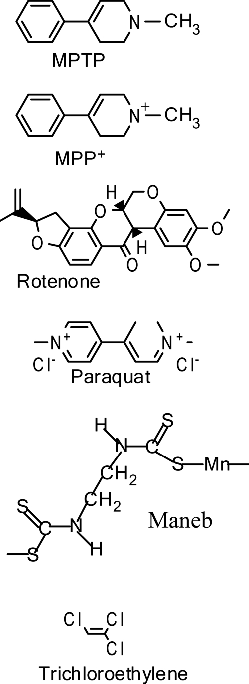
Recent advances: A new toxin model using the mitochondrial toxin trichloroethylene was recently described and novel methods, such as intranasal exposure to toxins, have been explored. Additionally, recent research conducted in toxin models of parkinsonism provides an emerging emphasis on extranigral aspects of PD pathology.

Critical issues: Unfortunately, none of the existing animal models of experimental PD completely mimics the etiology, progression, and pathology of human PD.

Future directions: Continued efforts to optimize established animal models of parkinsonism, as well as the development and characterization of new animal models are essential, as there still remains a disconnect in terms of translating mechanistic observations in animal models of experimental PD into bona fide disease-modifying therapeutics for human PD patients.


Figures

FIG. 1.
FIG. 1.
Dysregulated mitochondrial dynamics in Parkinson's disease toxin models. Dynamic balance between mitochondrial fission and fusion is required for normal cell homeostasis and function. Mitochondrial fusion occurs when mitofusion proteins (Mfn) link the outer mitochondrial membranes of two separate mitochondria, and the protein Opa1, which resides in the inner mitochondrial membrane, facilitates fusion of the inner mitochondrial membranes, resulting in the fusion of one mitochondria from two. Mitochondrial fission occurs when the fission protein Fis1 demarks the outer mitochondrial membrane, and by interaction with Drp1, promotes the fission of a single mitochondria into two individual mitochondria. Excessive mitochondria fusion is detrimental to cell survival, and rotenone promotes aberrant mitochondrial fusion.
FIG. 2.
FIG. 2.
Chemical structures of toxins that induce mitochondrial dysfunction and are used to model experimental Parkinson's disease.
FIG. 3.
FIG. 3.
Schematic of the MPTP/MPP+ metabolic pathway and neurotoxicity. Following systemic administration, MPTP readily crosses the blood-brain barrier (BBB). Within the brain, MPTP is metabolized into the toxic MPP+ species by the enzymatic action of monoamine oxidase-b (MAO-B) within glia cells. The MPP+ is then released into the extracellular space where it is selectively taken up into dopamine neurons by the dopamine transporter (DAT). Once inside the DAergic neurons, the MPP+ is taken up by and concentrated within mitochondria, where it binds and inhibits mitochondrial complex I (C I), inducing general mitochondrial dysfunction and generating oxidative stress, culminating in cell death.
FIG. 4.
FIG. 4.
Mitochondrial dysfunction and the oxidative stress model of dopamine neuron cell death. Within DA neurons, the toxins MPP+ and rotenone inhibit mitochondrial complex I (c-I), decreasing endogenous antioxidants and generating oxidative stress from complexes I and III (c I, c III, respectively) which leads to oxidation of macromolecules. Additionally, cytochrome c (Cyt c) is released from the intermitochondrial space, activating caspase signaling and subsequent apoptotic cell death. The toxin paraquat accepts electrons from complex I (c I) and functions as a redox cycler to generate oxidative stress and subsequent mitochondrial dysfunction leading to DA neuron death in a manner similar to MPP+ and rotenone. The toxin maneb (MB) is believed to induce DA neuron cell death via mechanisms similar to MPP+, rotenone and PQ, although MB does so by inhibiting mitochondrial complex III (c-III).
FIG. 5.
FIG. 5.
Schematic of oxidative phosphorylation and the mitochondrial electron transport chain. The electron transport chain resides within the inner mitochondrial membrane (IM) and provides cellular energy by oxidative phosphorylation. The toxins MPP+ and rotenone inhibit complex I (c I), while the toxin maneb (MB) is an inhibitor of complex III (c III). As a result of inhibition, oxidative stress is generated by C I and C III. C II, complex II; c IV, complex IV; c V, complex V; IMS, intermembrane space; OM, outer mitochondrial membrane.
FIG. 6.
FIG. 6.
Mitochondrial dysfunction and the ATP energy crisis model of dopamine neuron cell death. Inhibition of mitochondrial complex I (c I) and redox cycling by paraquat (PQ) decreases ATP levels, which may lead to compromised function of the ubiquitin proteasome system (UPS) and bioenergetic crisis within DA neurons, culminating in cell death.
FIG. 7.
FIG. 7.
Mitochondrial dysfunction and the mitochondrial calcium overload/glutamatergic excitotoxic cell death model in dopamine neurons. Within DA neurons, rotenone treatment causes an increase in glutamate and a cytoplasmic influx of calcium (Ca2+) through the NMDA receptor (NMDA R) and through cav1.3 L-type Ca2+ channels, likely culminating in excitotoxic cell death. Paraquat (PQ) may also contribute to excitotoxic DA neuron death due to its function as a redox cycler.
FIG. 8.
FIG. 8.
The redox cycling mechanism of paraquat. The paraquat dication (PQ2+) accepts electrons (e-) from complex I (c I) and is reduced, generating the paraquat radical (PQ+) which potently reacts with O2 to generate superoxide (O•-).

References

    1. Ahmadi FA. Linseman DA. Grammatopoulos TN. Jones SM. Bouchard RJ. Freed CR. Heidenreich KA. Zawada WM. The pesticide rotenone induces caspase-3-mediated apoptosis in ventral mesencephalic dopaminergic neurons. J Neurochem. 2003;87:914–921. - PubMed
    1. Anderson G. Noorian AR. Taylor G. Anitha M. Bernhard D. Srinivasan S. Greene JG. Loss of enteric dopaminergic neurons and associated changes in colon motility in an MPTP mouse model of Parkinson's disease. Exp Neurol. 2007;207:4–12. - PMC - PubMed
    1. Arnold B. Cassady SJ. VanLaar VS. Berman SB. Integrating multiple aspects of mitochondrial dynamics in neurons: Age-related differences and dynamic changes in a chronic rotenone model. Neurobiol Dis. 2011;41:189–200. - PMC - PubMed
    1. Ascherio A. Chen H. Weisskopf MG. O'Reilly E. McCullough ML. Calle EE. Schwarzschild MA. Thun MJ. Pesticide exposure and risk for Parkinson's disease. Ann Neurol. 2006;60:197–203. - PubMed
    1. Barrot M. Calza L. Pozza M. Le Moal M. Piazza PV. Differential calbindin-immunoreactivity in dopamine neurons projecting to the rat striatal complex. Eur J Neurosci. 2000;12:4578–4582. - PubMed

Publication types

MeSH terms

Substances

LinkOut - more resources