Review
. 2012 Jan;24(1):202-14.
doi: 10.1111/j.1365-2826.2011.02151.x.

Anabolic androgenic steroid abuse: multiple mechanisms of regulation of GABAergic synapses in neuroendocrine control regions of the rodent forebrain

Affiliations
Review

Anabolic androgenic steroid abuse: multiple mechanisms of regulation of GABAergic synapses in neuroendocrine control regions of the rodent forebrain

J G Oberlander et al. J Neuroendocrinol. 2012 Jan.

Erratum in

  • J Neuroendocrinol. 2012 May;24(5):849

Abstract

Anabolic androgenic steroids (AAS) are synthetic derivatives of testosterone originally developed for clinical purposes but are now predominantly taken at suprapharmacological levels as drugs of abuse. To date, almost 100 different AAS compounds that vary in metabolic fate and physiological effects have been designed and synthesised. Although they are administered for their ability to enhance muscle mass and performance, untoward side effects of AAS use include changes in reproductive and sexual behaviours. Specifically, AAS, depending on the type of compound administered, can delay or advance pubertal onset, lead to irregular oestrous cyclicity, diminish male and female sexual behaviours, and accelerate reproductive senescence. Numerous brains regions and neurotransmitter signalling systems are involved in the generation of these behaviours, and are potential targets for both chronic and acute actions of the AAS. However, critical to all of these behaviours is neurotransmission mediated by GABA(A) receptors within a nexus of interconnected forebrain regions that includes the medial preoptic area, the anteroventral periventricular nucleus and the arcuate nucleus of the hypothalamus. We review how exposure to AAS alters GABAergic transmission and neural activity within these forebrain regions, taking advantage of in vitro systems and both wild-type and genetically altered mouse strains, aiming to better understand how these synthetic steroids affect the neural systems that underlie the regulation of reproduction and the expression of sexual behaviours.


Figures

Figure 1
Figure 1
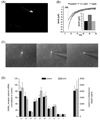
Properties of GnRH Neurones. (A) Representative photomicrograph of a GnRH neurone from the GFP-GnRH transgenic line generously provided by SM Moenter (University of Michigan; [95]). (B) Acute modulation of GABAA receptor-mediated sPSCs in a GnRH neurone by 17α-methyltestosterone. Average currents (>30 neurones under each condition) recorded in aCSF alone (control), in aCSF supplemented with 1 µM 17α-methyltestosterone (17α-MeT) and following return to aCSF (wash). (C) Photomicrographs illustrating harvesting of the contents of a fluorescent GFP-GnRH neurone for single cell PCR analysis. (D) GABAA receptor subunit mRNA levels in GnRH neurones of gonadally-intact oil-injected (control) and AAS-treated male mice. Data are presented as the 2−ΔCT values, which indicate the average levels (relative to the housekeeping gene β-actin) of subunit mRNAs in GnRH neurones isolated from control (black; n= 10 mice) and AAS-treated (grey; n = 10 mice) for analysis of GABAA receptor subunit mRNA levels and from a separate cohort of 7 control and 7 AAS-treated mice for analysis of GnRH mRNA levels. Data in (D) are from Penatti et al. [69].
Figure 2
Figure 2
Model of AAS Action in Adolescent Male Mice Chronically Treated with 17α-Methyltestosterone. Gonadally-intact male mice were treated for 4 weeks beginning at postnatal day (PN) 25–28 with 7.5 mg/kg/day for 6 days/week or with oil (control). (A) Model schematic of GABAergic mPOA inputs to GnRH neurones. Electrophysiological assessments made following the treatment period indicated that AAS treatment (B) significantly (p = 0.015) increased action potential frequency in mPOA neurones of AAS-treated (grey; n = 58 neurones) versus control (black; n = 72 neurones) mice and promoted a trend towards a decrease in the number of neurones that displayed irregular firing patterns; (C) had no effect on the amplitude or kinetics of GABAA receptor-mediated sPSCs recorded from GnRH neurones, as illustrated by averaged currents from neurones from control (grey) and AAS-treated (black) mice, but did significantly (p = 0.014) increase the frequency of these sPSCs (n = 12 neurones for control and n = 13 neurones for AAS-treated); and (D) significantly (p = 2.03 × 10⁻⁴) decreased the frequency of action potentials in GnRH neurones from AAS-treated (grey; n = 18 cells) versus control (black; n = 22 cells) without altering action potential patterning in these GnRH neurones. Data are modified from Penatti et al [69].
Figure 3
Figure 3
AAS Antagonism of Endogenous ER Signalling in the AR-deficient Tfm Mouse. (A) Graphical representation of averaged peak sIPSC amplitude from Tfm mice chronically treated with oil alone (Control), the AAS mixture in oil (AAS), tamoxifen (Tx), the AAS cocktail and tamoxifen (Tx+AAS), formestane (Frm) the AAS cocktail and formestane (Frm+AAS), 17β-oestradiol and the AAS cocktail (E2+AAS) or 17β-oestradiol alone (E2). Identical letters indicate means that were not statistically different from one another as assessed by two-way ANOVA followed by the means comparison by least significant means. Numbers in parentheses indicate numbers of cells. (B) Means ± standard errors of the mean levels of testosterone (T) and 17β-oestradiol (E2) in mPOA tissue harvested from control (n = 6 mice for T; n = 8 mice for E2) or AAS-injected (n = 4 mice for T; n = 8 mice for E2) Tfm male mice. Data are modified from Penatti et al. [84].
Figure 4
Figure 4
Characteristics of Neuroendocrine Control Neurones in the GT wild type and GT AR-mutant mouse line. GT AR-mutant and wild type mice were identified by a TaqMan®SNP genotyping assay, which consisted of a common forward and reverse primer and two unique probes based on wild type versus mutant (single base deletion) sequences coupled with different fluorophores (forward primer: 5’-AACTTTCCGCTGGCTCTGT-3’; reverse primer: 5’-CTTGATACGGGCGTGTGGAT-3’; wild type probe: VIC – ACCCCCCGCCCCCT; mutant probe: 6FAM-CACCCCCGCCCCCT). (A) Average AP frequencies recorded in the on-cell configuration from GnRH, mPOA and AVPV neurones. ** Indicates values in wile type male mice were significantly different from AR-mutants with p < 0.01. (B) Relative levels of mRNA encoding ERα and ERβ in mPOA tissue from GT AR mutant and wild type male mice. ^ Indicates a trend that did not attain significance (p = 0.059) towards higher levels of ERα in AR-mutant than wild type male mice. (C) Steady state levels of kisspeptin mRNA were dramatically lower in the mPOA and dramatically elevated in the arcuate of GT AR-mutant versus wild type mice (*** indicates p < 0.001). (D) Immunohistochemical assessment with kisspeptin-10 antibody (Chemicon AB9754, 1:1000) demonstrated significant differences (*** indicates p < 0.001) in the numbers of immunopositive neurones within the 50 µm directly adjacent to the edge of the ventricle in the AVPV and in the arcuate that mirrored differences in mRNA levels for these two regions in GT AR-mutant versus wild type mice. (E) Treatment for two weeks of GT ARmutant male mice with 0.1 mg/kg 17β-oestradiol (E2) restored the pattern of kisspeptin mRNA expression in both the mPOA and the arcuate observed in wild type GT mice.

Similar articles

Cited by

References

    1. Basaria S, Wahlstrom JT, Dobs AS. Clinical review 138: Anabolic-androgenic steroid therapy in the treatment of chronic diseases. J Clin Endocrinol Metab. 2001;86:5108–5117. - PubMed
    1. Shahidi NT. A review of the chemistry, biological action, and clinical applications of anabolic-androgenic steroids. Clin Ther. 2001;23:1355–1390. - PubMed
    1. Kochakian C, Yesalis CE. Anabolic-androgenic steroids: a historical perspective and definition. In: Yesalis CE, editor. Anabolic Steroids in Sport and Exercise. Champaign: Human Kinetics; 2000. pp. 4–33.
    1. Llewellyn W. Body of Science. 6th Edition. Jupiter, FL: 2007. Anabolics; pp. vii–ix.
    1. Trenton AJ, Currier GW. Behavioural manifestations of anabolic steroid use. CNS Drugs. 2005;19:571–595. - PubMed

Publication types