Review
. 2011 Dec;278(23):4486-96.
doi: 10.1111/j.1742-4658.2011.08166.x. Epub 2011 May 31.

Unique ganglioside binding by botulinum neurotoxins C and D-SA

Affiliations
Review

Unique ganglioside binding by botulinum neurotoxins C and D-SA

Abby R Kroken et al. FEBS J. 2011 Dec.

Abstract

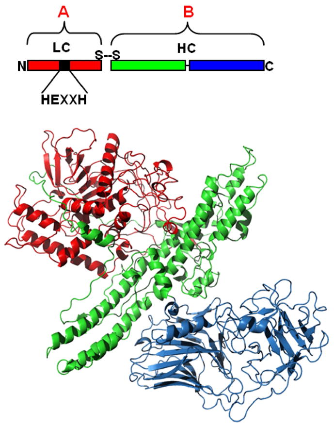
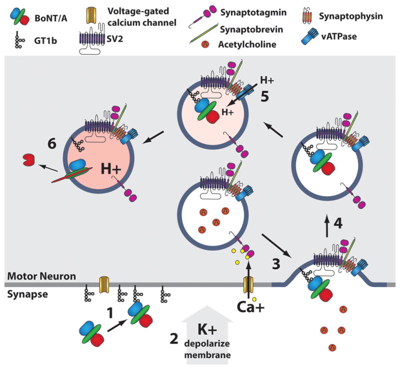
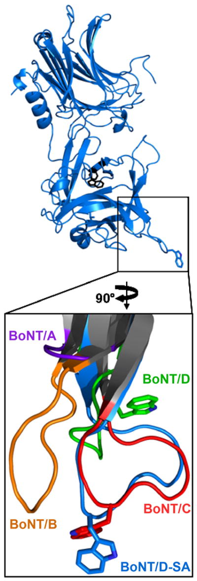
The botulinum neurotoxins (BoNTs) are the most potent protein toxins for humans. There are seven serotypes of BoNTs (A-G), based on a lack of cross-antiserum neutralization. The BoNT/C and BoNT/D serotypes include mosaic toxins that are organized as D-C and C-D toxins. One BoNT D-C mosaic toxin, BoNT/D-South Africa (BoNT/D-SA), was not fully neutralized by immunization with a vaccine composed of either prototype BoNT/C-Stockholm or BoNT/D-1873. Whereas several BoNT serotypes utilize dual receptors (gangliosides and proteins) to bind to and enter neurons, the basis for BoNT/C and BoNT/D entry into neurons is less well understood. Recent studies solved the crystal structures of the receptor-binding domains of BoNT/C, BoNT/D, and BoNT/D-SA. Comparative structural analysis showed that BoNT/C, BoNT/D and BoNT/D-SA lacked components of the ganglioside-binding pocket that exists within other BoNT serotypes. With the use of structure-based alignments, biochemical analyses, and cell-binding approaches, BoNT/C and BoNT/D-SA have been shown to possess a unique ganglioside-binding domain, the ganglioside-binding loop. Defining how BoNTs enter host cells provides insights towards understanding the evolution and extending the potential therapeutic and immunological values of the BoNT serotypes.


Figures

Figure 1
Figure 1. Structure-Function Organization of the Botulinum Neurotoxins
(Upper Panel) BoNTs are AB toxins composed of independent functional domains linked by a disulphide bond. The N-terminal light chain (LC, Red) encodes the enzymatic domain, while the heavy chain (HC) encodes two independent domains, the receptor binding domain (HCR, Blue) and the translocation domain (HCT, Green). (Lower Panel) Crystal structure of BoNT/A showed three functional domains: LC (Red), (HCR (Blue), and HCT (Green). PDB:3BTA; solved by Lacy and Stevens [4].
Figure 2
Figure 2. Entry of BoNTs into Neurons
Several BoNT serotypes enter neurons upon membrane depolarization, using BoNT/A several steps that can be resolved include: Step 1 HCR of BoNT/A binds GT1b on the plasma membrane of unstimulated neuron (blue). Step 2 Membrane depolarization, elicited in cultured cells using elevated extracellular potassium, triggers the opening of voltage-gated calcium channels, allowing influx of calcium. Step 3 Intracellular calcium binds synaptotagmin I/II, located in isolation and in complex with SV2, which signals for fusion of synaptic vesicles to the plasma membrane. Vesicle fusion exposes L4 loop of SV2, the protein receptor for BoNT/A. The HCR binds GT1b and SV2 simultaneously. Step 4 Complexes of synaptic vesicle proteins are endocytosed to be recycled. Step 5 The vacuolar ATPase acidifies the lumen of the synaptic vesicle. Step 6 The acidic environment triggers insertion of the HCT domain which facilitates translocation of a partially unfolded LC (red) through a channel made by HCT (green). Once in the cytoplasm, LC cleaves SNAP-25.
Figure 3
Figure 3. Crystal structures of HCR/C, HCR/D, and HCR/D-SA
Shown are overlays of the crystal structure of the HCR/D-SA (blue) with HCR/C (left panel, red) RMSD: 0.46 Δ and HCR/D-SA (blue) with HCR/D (right panel, green) RMSD: 2.47Δ. PDB: HCR/C, 3N7K; HCR/D, 3N7J; HCR/D-SA, 3N7L. Reproduced from [62] with permission.
Figure 4
Figure 4. Ganglioside Binding Pocket of HCR/A overlay with HCR/D-SA
HCR/D-SA (blue) includes conserved internal Phe1280 and Trp1282 (grey), and corresponding residues that represent the Ganglioside Binding Pocket of HCR/A (green). Enlarged views of the Ganglioside Binding Pocket of HCR/A (lower) and the corresponding region of HCR/D-SA (upper) are shown. Residues that contribute to ganglioside binding of HCR/A (Glu1203, His1253, Ser1264, Trp1266 and Tyr1267) and corresponding residues within HCR/D-SA are shown. Reproduced from [62] with permission.
Figure 5
Figure 5. Ganglioside Binding Loops of HCR/C, HCR/D, and HCR/D-SA overlay with HCR/A and HCR/B
HCR/D-SA (blue, upper) includes conserved Phe1280 and Trp1282 (black), and the Ganglioside Binding Loop (GBL) is enlarged, rotated, (lower) and aligned with the structurally analogous β-hairpin loops of BoNT/A (purple), /B (orange), /C (red), /D (green). HCR/C and /D-SA loops. Note, BoNT/B has an extended β-hairpin loop like HCR/C, HCR/D, and HCR/D-SA, but lacks a tryptophan residue. BoNT/A, in contrast, does not have an extended β-hairpin loop. Reproduced from [62] with permission.
Figure 6
Figure 6. Alignment of HCR/C to the HCR/B-Synaptotagmin complex
A. Crystal structure of HCR/B (green) bound to synaptotagmin peptide (grey, PBD: 2NM1 [26], aligned with HCR/C (red, PDB: 3N7K). Trp1266 and Tyr1267 of the ganglioside binding pocket (GBP) are shown in cyan. The ganglioside binding loop (GBL1) of HCR/C is in purple with Trp1258 shown. Structures were aligned so that Trp1266 of HCR/B and Trp1258 of HCR/C are parallel with the plasma membrane (dashed line, PM). B. The 1250 loop described by Stevens and coworkers [64] potentially penetrates the PM while the synaptotagmin peptide fits into a crevice within the C terminus of HCR/B. C. Same alignment as in A. except HCR/B was omitted for clarity. Tyr1273 maps to the GBP and is shown in cyan. Trp1258 is positioned to interact with PM embedded ganglioside and along with Tyr1259 may penetrate the PM lipid bilayer, with another loop, GBL2, also membrane associated.

Similar articles

Cited by

References

    1. Gill DM. Bacterial toxins: a table of lethal amounts. Microbiol Mol Biol Rev. 1982;46:86–94. - PMC - PubMed
    1. Singh BR, Gimenez JA, DasGupta BR. Comparative molecular topography of botulinum neurotoxins from Clostridium butyricum and Clostridium botulinum type E. Biochim Biophys Acta. 1991;1077:119–126. [pii] - PubMed
    1. Hill KK, Smith TJ, Helma CH, Ticknor LO, Foley BT, Svensson RT, Brown JL, Johnson EA, Smith LA, Okinaka RT, et al. Genetic diversity among Botulinum Neurotoxin-producing clostridial strains. J Bacteriol. 2007;189:818–832. doi: 10.1128/JB.01180-06. JB.01180-06 [pii] - DOI - PMC - PubMed
    1. Lacy DB, Tepp W, Cohen AC, DasGupta BR, Stevens RC. Crystal structure of botulinum neurotoxin type A and implications for toxicity. Nat Struct Mol Biol. 1998;5:898–902. - PubMed
    1. Breidenbach MA, Brunger AT. Substrate recognition strategy for botulinum neurotoxin serotype A. Nature. 2004;432:925–929. doi: 10.1038/nature03123. nature03123 [pii] - DOI - PubMed

Publication types