. 2011 May 9:11:13.
doi: 10.1186/1472-6785-11-13.

Behavioral mechanisms and morphological symptoms of zombie ants dying from fungal infection

Affiliations

Behavioral mechanisms and morphological symptoms of zombie ants dying from fungal infection

David P Hughes et al. BMC Ecol. .

Abstract

Background: Parasites that manipulate host behavior can provide prominent examples of extended phenotypes: parasite genomes controlling host behavior. Here we focus on one of the most dramatic examples of behavioral manipulation, the death grip of ants infected by Ophiocordyceps fungi. We studied the interaction between O. unilateralis s.l. and its host ant Camponotus leonardi in a Thai rainforest, where infected ants descend from their canopy nests down to understory vegetation to bite into abaxial leaf veins before dying. Host mortality is concentrated in patches (graveyards) where ants die on sapling leaves ca. 25 cm above the soil surface where conditions for parasite development are optimal. Here we address whether the sequence of ant behaviors leading to the final death grip can also be interpreted as parasite adaptations and describe some of the morphological changes inside the heads of infected workers that mediate the expression of the death grip phenotype.

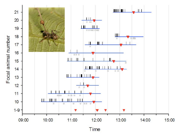
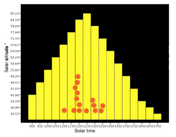
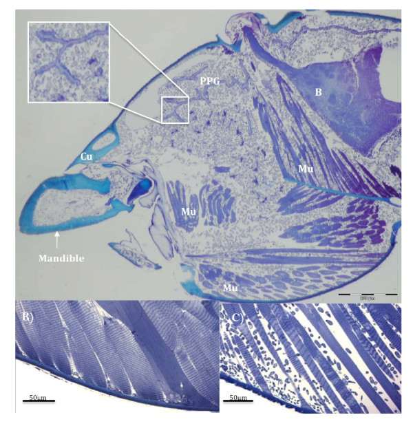
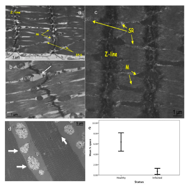
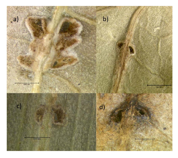
Results: We found that infected ants behave as zombies and display predictable stereotypical behaviors of random rather than directional walking, and of repeated convulsions that make them fall down and thus precludes returning to the canopy. Transitions from erratic wandering to death grips on a leaf vein were abrupt and synchronized around solar noon. We show that the mandibles of ants penetrate deeply into vein tissue and that this is accompanied by extensive atrophy of the mandibular muscles. This lock-jaw means the ant will remain attached to the leaf after death. We further present histological data to show that a high density of single celled stages of the parasite within the head capsule of dying ants are likely to be responsible for this muscular atrophy.

Conclusions: Extended phenotypes in ants induced by fungal infections are a complex example of behavioral manipulation requiring coordinated changes of host behavior and morphology. Future work should address the genetic basis of such extended phenotypes.


Figures

Figure 1
Figure 1
Zombie ant behavior. Focal animal observation periodogram of ants infected by Ophiocordyceps unilateralis s.l. The blue horizontal bars mark the observation period, the red triangles mark moment of biting, the vertical bars mark spasm events and the grey diamonds the falling off events. For four individuals that belong to focal animals 1-9 only the biting time was recorded. The biting time was recorded for 16 ants but only 15 triangles are visible as two ants bite at exactly the same time (12:05). Inset picture shows a dead ant on a leaf with the fungal stalk and spore body that emerged from the head.
Figure 2
Figure 2
Synchronized manipulation of ants by fungi. A sun position chart of the death grip. Solar altitude is represented by the yellow bars and plotted against the y-axis and the biting times are the red circles and plotted on the x-axis in solar time (this is true local time accounting for longitude and different from Time in Figure 1). The red circles are stacked to prevent overlapping. At 11:47 two ants bite so only 15 circles are visible though 16 ants were recorded.
Figure 3
Figure 3
Heads of manipulated ants colonized by fungi. A (top panel) is a light micrograph (LM) saggital section through the head of an O. unilateralis s.l infected ant that was biting a leaf at the moment of fixation (i.e. alive). The small grey blobs are fungal hyphal bodies that fill the head and mandibles. Note the spacing between the muscle fibers. The insect shows a close up of hyphal bodies around the post-pharyngeal gland (PPG). B is the brain, Mu, Muscles and Cu is cuticle. B) is a LM of healthy muscle and C) is a LM of muscle from a behaviorally manipulated ant that was biting a leaf and alive when removed for fixation. The small blobs between the fibers are fungal cells.
Figure 4
Figure 4
Muscular atrophy in O. unilateralis s.l infected ants. A) and B) are transmission electron micrographs (TEM) of infected ant mandible muscles. C) TEM shows uninfected muscles. The interfibrillar spaces (IFS) and reduced mitochondria (M) are evident in A & B. The z-line is thinned in A) and in B) an example of a broken z-line is shown by the white arrow. C) shows healthy muscle with prominent sarcoplasmic reticula (SR) and mitochondria. D) is a TEM of the mandible muscle of an O. unilateralis infected ant. The fibers are stretched and tearing is evident as lines running perpendicular to the sarcomeres. The large irregular structures between the muscle fibers are fungal cells. Note the impressions (white arrow) formed where they meet the muscle. The internal structures of the fungal cells have not been preserved using our method and the large white area within these cells reflects that and is therefore an artefact of fixing. E) Quantification of atrophy (mean (+/- SE) percent of interfibrillar space in the muscles cells) in healthy and infected ants. The difference is significant (see text).
Figure 5
Figure 5
Damage to plant tissue by zombie ants. Photographs of abaxial vein and leaf lamina where zombie ants bit leading to scar formation by the plant. In A) two large holes are visible where the mandibles penetrated the lamina. In addition smaller marks are evident that formed as a result of rasping before the final death grip (see text). In B-D only one set of mandible marks occur. In all cases the vein has been cut. The arrows bars are 0.4 mm, 0.8 mm, 0.6 mm and 0.5 mm for A,B,C,D respectively.

Similar articles

Cited by

References

    1. Moore J. Parasites and the behavior of animals. Oxford: Oxford University Press; 2002.
    1. Lafferty K, Kuris AM. Parasitic castration: the evolution and ecology of body snatchers. Trends in Parasitology. 2009;25(12):564–572. doi: 10.1016/j.pt.2009.09.003. - DOI - PubMed
    1. Poulin R. Progenesis and reduced virulence as an alternative transmission strategy in a parasitic trematode. Parasitology. 2001;123:623–630. - PubMed
    1. Lefevre T, Adamo SA, Biron DG, Misse D, Hughes D, Thomas F. Invasion of the Body Snatchers: the diversity and evolution of manipulative strategies in host-parasite interactions. Advances in Parasitology. 2009;68:45–83. - PubMed
    1. Dawkins R. The extended phenotype. Oxford: W.H. Freeman; 1982.

Publication types

LinkOut - more resources