. 2011 May 9:10:70.
doi: 10.1186/1476-511X-10-70.

S1P, dihydro-S1P and C24:1-ceramide levels in the HDL-containing fraction of serum inversely correlate with occurrence of ischemic heart disease

Affiliations

S1P, dihydro-S1P and C24:1-ceramide levels in the HDL-containing fraction of serum inversely correlate with occurrence of ischemic heart disease

Kelley M Argraves et al. Lipids Health Dis. .

Abstract

Background: The lysosphingolipid sphingosine 1-phosphate (S1P) is carried in the blood in association with lipoproteins, predominantly high density lipoproteins (HDL). Emerging evidence suggests that many of the effects of HDL on cardiovascular function may be attributable to its S1P cargo.

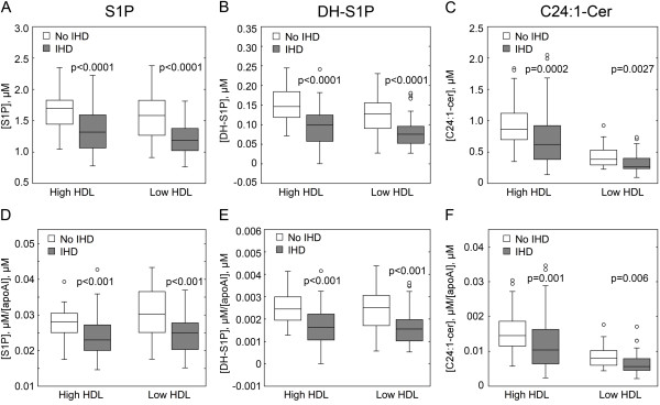
Methods: Here we have evaluated how levels of S1P and related sphingolipids in an HDL-containing fraction of human serum correlate with occurrence of ischemic heart disease (IHD). To accomplish this we used liquid chromatography-mass spectrometry to measure S1P levels in the HDL-containing fraction of serum (depleted of LDL and VLDL) from 204 subjects in the Copenhagen City Heart Study (CCHS). The study group consisted of individuals having high serum HDL cholesterol (HDL-C) (females:≥ 73.5 mg/dL; males:≥ 61.9 mg/dL) and verified IHD; subjects with high HDL-C and no IHD; individuals with low HDL-C (females:≤ 38.7 mg/dL; males:≤ 34.1 mg/dL) and IHD, and subjects with low HDL-C and no IHD.

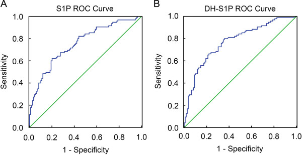
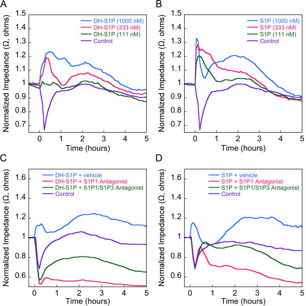
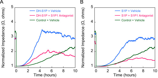
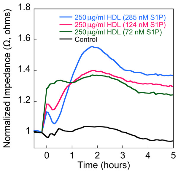
Results: The results show a highly significant inverse relationship between the level of S1P in the HDL-containing fraction of serum and the occurrence of IHD. Furthermore, an inverse relationship with IHD was also observed for two other sphingolipids, dihydro-S1P and C24:1-ceramide, in the HDL-containing fraction of serum. Additionally, we demonstrated that the amount of S1P on HDL correlates with the magnitude of HDL-induced endothelial cell barrier signaling.

Conclusions: These findings indicate that compositional differences of sphingolipids in the HDL-containing fraction of human serum are related to the occurrence of IHD, and may contribute to the putative protective role of HDL in IHD.


Figures

Figure 1
Figure 1
An inverse correlation exists between the occurrence of IHD and levels of S1P and DH-S1P in HDL-containing fractions from CCHS subject serum. S1P (A) DH-S1P (B) and C24.1 ceramide (C) levels were measured by blinded LC-MS-MS analysis of 55 HDL-containing preparations from CCHS individuals with high HDL-C and no evidence of IHD, 53 samples from individuals with high HDL-C and evidence of IHD, 54 samples from individuals with low HDL-C and no evidence of IHD and 42 samples from individuals with low HDL-C and evidence of IHD. In D-F, levels of S1P and DH-S1P in HDL-containing serum fractions from CCHS individuals with and without IHD are assessed relative to apoA-I content. Levels of apoA-I in samples were quantified by immunoturbidometric assay using a Cobas Fara analyzer. Indicated p-values are derived from a Tukey's comparison following a one-way ANOVA test conducted at level of significance 0.05. Data are presented in box-and-whisker diagrams. For the box-and-whisker diagrams, the boxes correspond to the interquartile range (IQR). The horizontal bar within the box is drawn at the height of the median. The whiskers indicate the range of the data within 1.5 X IQR with outliers indicated as circles.
Figure 2
Figure 2
Receiver operating characteristic (ROC) curves for the ability of S1P and DH-S1P to distinguish between subjects with and without IHD. Smaller values of the corresponding measure indicate stronger evidence for the presence of IHD. ROC curves were constructed using SPSS v16.
Figure 3
Figure 3
DH-S1P enhances endothelial barrier in an S1P1 dependent manner. Confluent EC monolayers were grown under serum-free conditions until a minimal TEER plateau was reached. In A, EC monolayers were incubated with varying concentrations of DH-S1P [111-1000 nM]. In B, EC monolayers were incubated with varying concentrations of S1P [111-1000 nM]. In C, EC monolayers were incubated with DH-S1P [1000 nM] plus and minus the S1P1 antagonist W146 or the S1P1/S1P3 antagonist VPC23019 (each at 10 μM). In D, EC monolayers were incubated with S1P [1000 nM] plus and minus the S1P1 antagonist W146 or the S1P1/S1P3 antagonist VPC23019 (each at 10 μM). Each of the TEER tracings shown is an average from three independent experiments each with two replicates per condition. Impedance values were normalized by dividing each value by the level of impedance measured just prior to the addition of effectors. As a control for A and B, monolayers were treated with 40 μg/ml delipidated albumin (Control), a concentration corresponding to the amount of BSA carrier used for the highest concentration of DH-S1P and S1P tested. As controls for C and D, ECs were treated with delipidated albumin-containing serum free medium (SFM) plus vehicle buffer.
Figure 4
Figure 4
DH-S1P enhances endothelial cell motility in an S1P1 dependent manner. EC monolayers were wounded with a burst of high electrical current as described previously [5] and the culture medium then supplemented with DH-S1P (1 μM) (A), or S1P (1 μM) (B) in the presence or absence of the S1P1 antagonist W146 (10 μM in DMSO vehicle). The migration of cells into the wounded areas was measured in real-time by electrical impedance. As a control in both experiments the medium was supplemented with delipidated BSA (Control) in PBS. Electrical impedance data are normalized to baseline following wounding. The data depicted are representative of two independent experiments and traces represent averages of two replicates per condition.
Figure 5
Figure 5
Levels of HDL-associated S1P dictate the magnitude of the TEER response. Confluent EC monolayers were incubated with native HDL (72 nM S1P) or HDL containing varying amounts of exogenously added S1P. HDL was added to achieve a final concentration of 250 μg protein/ml. As a control, monolayers were incubated with the vehicle buffer, 0.03 mM EDTA in Dulbecco's PBS (Control). Each of the TEER tracings shown is an average from two replicates per treatment. Impedance values were normalized by dividing each value by the level of impedance measured just prior to the addition of effectors. The results depicted are representative of two independent experiments.

References

    1. Yatomi Y, Igarashi Y, Yang L, Hisano N, Qi R, Asazuma N, Satoh K, Ozaki Y, Kume S. Sphingosine 1-phosphate, a bioactive sphingolipid abundantly stored in platelets, is a normal constituent of human plasma and serum. Journal of biochemistry. 1997;121(5):969–973. - PubMed
    1. Murata N, Sato K, Kon J, Tomura H, Yanagita M, Kuwabara A, Ui M, Okajima F. Interaction of sphingosine 1-phosphate with plasma components, including lipoproteins, regulates the lipid receptor-mediated actions. Biochem J. 2000;352(Pt 3):809–815. - PMC - PubMed
    1. Argraves KM, Argraves WS. HDL serves as an S1P signaling platform mediating a multitude of cardiovascular effects. J Lipid Res. 2007;48(11):2325–2333. - PubMed
    1. Sattler K, Levkau B. Sphingosine-1-phosphate as a mediator of high-density lipoprotein effects in cardiovascular protection. Cardiovasc Res. 2009;82(2):201–211. - PubMed
    1. Argraves KM, Gazzolo PJ, Groh EM, Wilkerson BA, Matsuura BS, Twal WO, Hammad SM, Argraves WS. High density lipoprotein-associated sphingosine 1-phosphate promotes endothelial barrier function. J Biol Chem. 2008;283(36):25074–25081. - PMC - PubMed

Publication types

MeSH terms

LinkOut - more resources