. 2011 May 9;9(1):24.
doi: 10.1186/1477-5956-9-24.

Evaluation of three high abundance protein depletion kits for umbilical cord serum proteomics

Affiliations

Evaluation of three high abundance protein depletion kits for umbilical cord serum proteomics

Bin Liu et al. Proteome Sci. .

Abstract

Background: High abundance protein depletion is a major challenge in the study of serum/plasma proteomics. Prior to this study, most commercially available kits for depletion of highly abundant proteins had only been tested and evaluated in adult serum/plasma, while the depletion efficiency on umbilical cord serum/plasma had not been clarified. Structural differences between some adult and fetal proteins (such as albumin) make it likely that depletion approaches for adult and umbilical cord serum/plasma will be variable. Therefore, the primary purposes of the present study are to investigate the efficiencies of several commonly-used commercial kits during high abundance protein depletion from umbilical cord serum and to determine which kit yields the most effective and reproducible results for further proteomics research on umbilical cord serum.

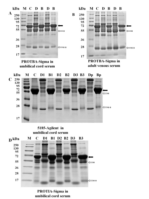
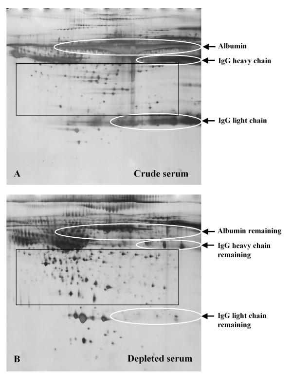
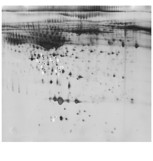
Results: The immunoaffinity based kits (PROTIA-Sigma and 5185-Agilent) displayed higher depletion efficiency than the immobilized dye based kit (PROTBA-Sigma) in umbilical cord serum samples. Both the PROTIA-Sigma and 5185-Agilent kit maintained high depletion efficiency when used three consecutive times. Depletion by the PROTIA-Sigma Kit improved 2DE gel quality by reducing smeared bands produced by the presence of high abundance proteins and increasing the intensity of other protein spots. During image analysis using the identical detection parameters, 411 ± 18 spots were detected in crude serum gels, while 757 ± 43 spots were detected in depleted serum gels. Eight spots unique to depleted serum gels were identified by MALDI- TOF/TOF MS, seven of which were low abundance proteins.

Conclusions: The immunoaffinity based kits exceeded the immobilized dye based kit in high abundance protein depletion of umbilical cord serum samples and dramatically improved 2DE gel quality for detection of trace biomarkers.


Figures

Figure 1
Figure 1
Comparison of depletion efficiency and repeatability of different kits on umbilical cord serum proteins samples by 1D-SDS-PAGE. Ten micrograms of crude, depleted or bound protein were loaded onto each lane and separated by 4-12% polyacrylamide gels. Proteins were visualized by Coomassie Blue staining. For examination of repeatability in umbilical cord serum depletion, the immunoaffinity columns (5185-Agilent and PROTIA-Sigma) were run three consecutive times with re-equilibration between depletion processes. However, for Blue Albumin and IgG Depletion Kit (PROTBA, Sigma-Aldrich, Saint Louis, MO, USA), serum samples were tested in two independent columns since its depletion efficiency was low even with a new column. A) Blue Albumin and IgG Depletion Kit (PROTBA, Sigma-Aldrich, Saint Louis, MO, USA) in umbilical cord serum depletion. B) Blue Albumin and IgG Depletion Kit (PROTBA, Sigma-Aldrich, Saint Louis, MO, USA) in adult venous serum depletion. C) Multiple Affinity Removal System (5185, Agilent, Santa Clara, CA, USA) in umbilical cord serum depletion. D) Immunoaffinity Albumin and IgG Depletion Kit (PROTIA, Sigma-Aldrich, Saint Louis, MO, USA) in umbilical cord serum depletion. Lanes: M: Prestained protein marker (Fermentas; 250, 130; 95, 72, 55, 36, 28, 17); C: Crude serum; D: Depleted serum; B: Bound proteins; D1-3: Depleted umbilical cord serum proteins of the first, second and third depletion process. B1-3: Bound proteins eluted from the column after the first, second and third depletion process. Dp: Depleted Proteins pooled by the three depletion processes. Bp: Bound proteins pooled by the three depletion processes Closed arrows: Protein band indicating albumin; Open arrows: Protein band indicating IgG; *heavy chain, and **light chain.
Figure 2
Figure 2
Two Dimensional Electrophoresis Gel image of crude and depleted umbilical cord serum samples. For each gel, 150 μg of protein was separated over pH range 4-7 (24 cm strips) and 12.5% SDS-polyacrylamide gel. The gel was visualized by silver staining. The Immunoaffinity Albumin and IgG Depletion Kit (PROTIA, Sigma-Aldrich, Saint Louis, MO, USA) Kit was used to deplete albumin and IgG in the umbilical cord serum. Removal of these proteins improved the resolution in the albumin and IgG areas on the gel and increased the intensity of low abundance proteins. A) Two Dimension Electrophoresis Gel image of crude umbilical cord serum. B) Two Dimension Electrophoresis Gel image of depleted umbilical cord serum by Immunoaffinity Albumin and IgG Depletion Kit (PROTIA, Sigma-Aldrich, Saint Louis, MO, USA). Circles: regions of gel containing albumin, heavy and light chain of IgG before (panel A) and after depletion (panel B). Rectangle: used to highlight different protein patterns in the two groups, indicating a large number of protein spots that emerged in the depleted gel.
Figure 3
Figure 3
Identification of protein spots unique to depleted umbilical cord serum gels by the Immunoaffinity Albumin and IgG Depletion kit (PROTIA, Sigma-Aldrich, Saint Louis, MO, USA). Circles: Ten spots unique to the depleted gels were selected for MALDI-TOF/TOF MS identification. Gel image is the same as in Fig 2B. The numbers correspond to the protein numbers in Table 2.

References

    1. Kyriakakou M, Malamitsi-Puchner A, Militsi H, Boutsikou T, Margeli A, Hassiakos D, Kanaka-Gantenbein C, Papassotiriou I, Mastorakos G. Leptin and adiponectin concentrations in intrauterine growth restricted and appropriate for gestational age fetuses, neonates, and their mothers. Eur J Endocrinol. 2008;158:343–348. doi: 10.1530/EJE-07-0692. - DOI - PubMed
    1. Anderson NL, Anderson NG. The human plasma proteome: history, character, and diagnostic prospects. Mol Cell Proteomics. 2002;1:845–867. doi: 10.1074/mcp.R200007-MCP200. - DOI - PubMed
    1. Song HJ, Zhang P, Guo XJ, Liao LM, Zhou ZM, Sha JH, Cui YG, Ji H, Liu JY. The proteomic analysis of human neonatal umbilical cord serum by mass spectrometry. Acta Pharmacol Sin. 2009;30:1550–1558. doi: 10.1038/aps.2009.140. - DOI - PMC - PubMed
    1. Page NM, Kemp CF, Butlin DJ, Lowry PJ. Placental peptides as markers of gestational disease. Reproduction. 2002;123:487–495. doi: 10.1530/rep.0.1230487. - DOI - PubMed
    1. Urbas L, Brne P, Gabor B, Barut M, Strlic M, Petric TC, Strancar A. Depletion of high-abundance proteins from human plasma using a combination of an affinity and pseudo-affinity column. J Chromatogr A. 2009;1216:2689–2694. doi: 10.1016/j.chroma.2008.10.104. - DOI - PubMed

LinkOut - more resources