. 2011 May 9:6:8.
doi: 10.1186/1748-7161-6-8.

Brace technology thematic series - The Sforzesco and Sibilla braces, and the SPoRT (Symmetric, Patient oriented, Rigid, Three-dimensional, active) concept

Affiliations

Brace technology thematic series - The Sforzesco and Sibilla braces, and the SPoRT (Symmetric, Patient oriented, Rigid, Three-dimensional, active) concept

Stefano Negrini et al. Scoliosis. .

Abstract

Background: Bracing is an effective strategy for scoliosis treatment, but there is no consensus on the best type of brace, nor on the way in which it should act on the spine to achieve good correction. The aim of this paper is to present the family of SPoRT (Symmetric, Patient-oriented, Rigid, Three-dimensional, active) braces: Sforzesco (the first introduced), Sibilla and Lapadula.

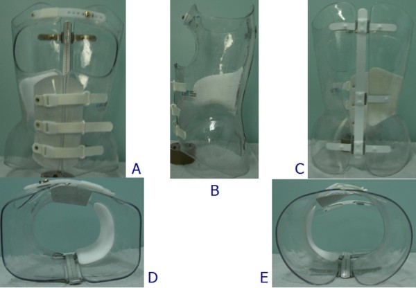
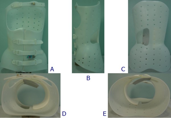
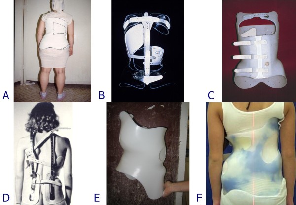
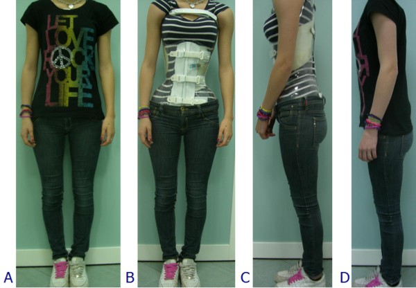
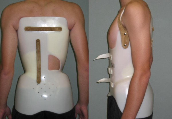
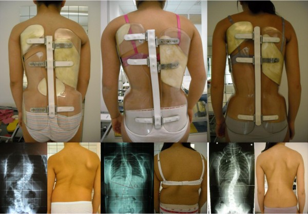
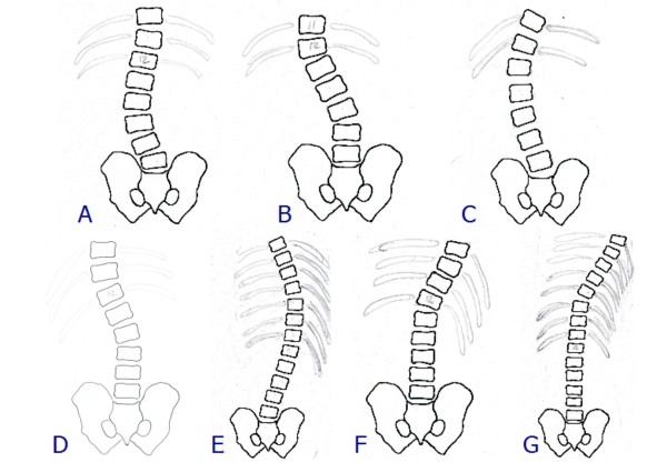
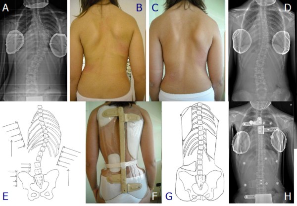
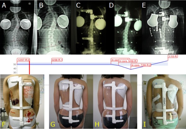
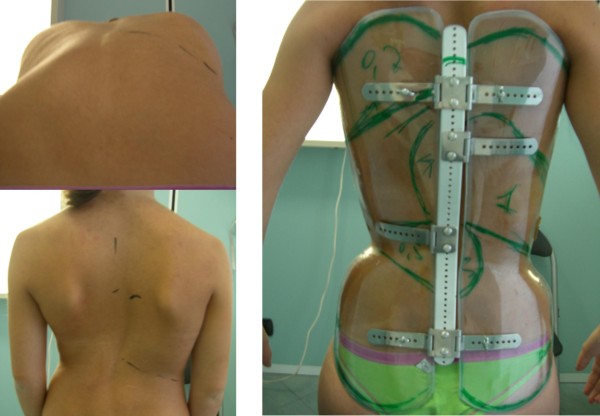
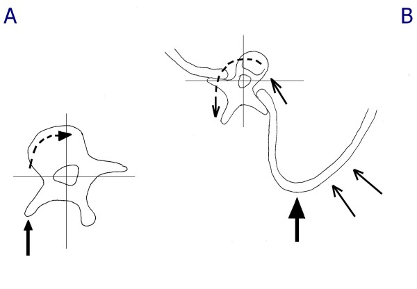
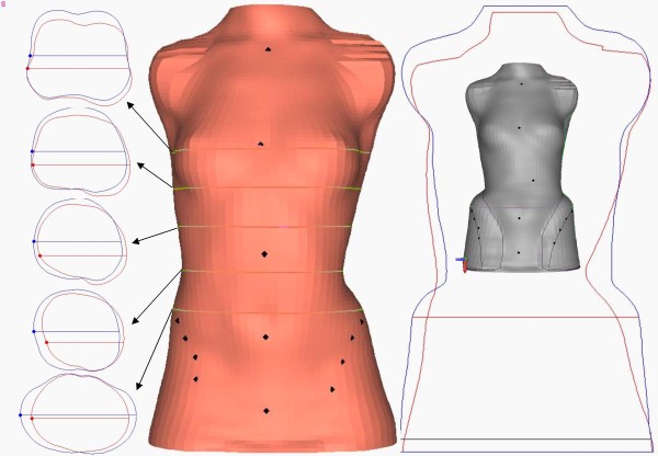
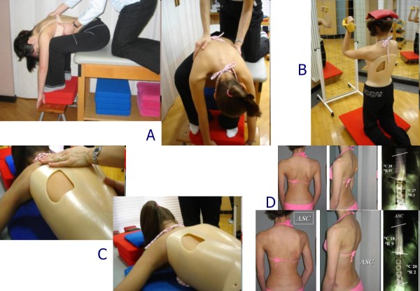
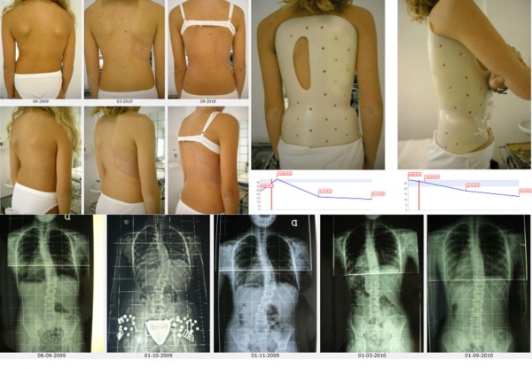
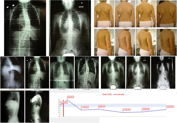
Methods: The Sforzesco brace was developed following specific principles of correction. Due to its overall symmetry, the brace provides space over pathological depressions and pushes over elevations. Correction is reached through construction of the envelope, pushes, escapes, stops, and drivers. The real novelty is the drivers, introduced for the first time with the Sforzesco brace; they allow to achieve the main action of the brace: a three-dimensional elongation pushing the spine in a down-up direction.Brace prescription is made plane by plane: frontal (on the "slopes", another novelty of this concept, i.e. the laterally flexed sections of the spine), horizontal, and sagittal. The brace is built modelling the trunk shape obtained either by a plaster cast mould or by CAD-CAM construction. Brace checking is essential, since SPoRT braces are adjustable and customisable according to each individual curve pattern.Treatment time and duration is individually tailored (18-23 hours per day until Risser 3, then gradual reduction). SEAS (Scientific Exercises Approach to Scoliosis) exercises are a key factor to achieve success.

Results: The Sforzesco brace has shown to be more effective than the Lyon brace (matched case/control), equally effective as the Risser plaster cast (prospective cohort with retrospective controls), more effective than the Risser cast + Lyon brace in treating curves over 45 degrees Cobb (prospective cohort), and is able to improve aesthetic appearance (prospective cohort).

Conclusions: The SPoRT concept of bracing (three-dimensional elongation pushing in a down-up direction) is different from the other corrective systems: 3-point, traction, postural, and movement-based. The Sforzesco brace, being comparable to casting, may be the best brace for the worst cases.


Figures

Figure 1
Figure 1
The Sforzesco brace. The Sforzesco brace: anterior (A), left (B), posterior (C), top (D), and bottom (E) views.
Figure 2
Figure 2
The Sibilla brace. The Sibilla brace: anterior (A), left (B), posterior (C), top (D), and bottom (E) views.
Figure 3
Figure 3
The Lapadula brace. The Lapadula brace: anterior (A), left (B), posterior (C), top (D), and bottom (E) views.
Figure 4
Figure 4
The Sforzesco brace owns its name to the Sforesco castles of Milan and Vigevano. The Sforzesco brace was named according to the two main cities of the experience of the main author (SN): Vigevano and Milano, which both have castles named Sforzesco for the Medieval Sforza family.
Figure 5
Figure 5
Braces at the base of the SPoRT concept development. The concept of SPoRT bracing was developed from the following braces: Risser cast (A), Lyon (B), Sibilla (C) and Milwaukee (D). The last changes made to the SPoRT braces also allowed us to consider among their ancestors the last Cheneau brace (E) and the Rigo Cheneau brace (F).
Figure 6
Figure 6
The Sforzesco brace invisibility. The Sforzesco brace was developed in a town of fashion (Milan), and some patients have stated that this is reflected in its design, that increases wearability.
Figure 7
Figure 7
Patients want correction and an invisible brace. These patients are wearing their braces in various everyday activities.
Figure 8
Figure 8
The Lapadula brace to treat scoliosis and hyperkyphosis. The Lapadula brace has much versatility and can be adapted to treat an hyperkyphosis associated with scoliosis.
Figure 9
Figure 9
The first Sforzesco brace causes a sudden lengthening of the trunk requiring correction in 2 months. Typical correction made to the first Sforzesco brace after a wearing period of 2 months in patients with important thoracic curves: it becomes too short under the concave shoulder and must be lengthen.
Figure 10
Figure 10
The slopes. The slopes. Low lumbar (A); High lumbar (B); Lumbar (C); Thoracolumbar (D); Thoracic (E); Distal thoracic (F); Proximal thoracic (G).
Figure 11
Figure 11
Example of correction on the frontal plane slopes. Example of correction on the frontal plane slopes of one patient with a thoracic curve of 48° (Risser 1). (A) X-ray pre-brace (48°); (B) aesthetics pre-brace; (C) aesthetics after 4 months of bracing; (D) x-ray without the brace after 4 months of bracing: corrected to 23° (i.e. reduction of 25°, -52%); (E) pre-brace planning with pushes on the right thoraco-lumbar and left thoracic slopes, as well as a stop to the right of the flanks; (F) constructed brace; (G) application of the pushes in the constructed brace; (H) in-brace x-ray with a correction to 13° (i.e. 35° of correction, 73%).
Figure 12
Figure 12
The Cheneauisation of the Sforzesco brace. We used the term Cheneauisation to underline that it derives from the contamination of our own brace with the internationally well-known Cheneau brace: in fact it aims at posturally changing the scoliosis curve through a thrust at level of the convexity of the apical proximal curve and an elevation/medialization of the shoulder at the concave side. Due to our own SPoRT principles the Cheneuization also includes an anteposition of this same shoulder. First line, from left to right: (A) x-rays of the patient at start of treatment (12/07), (B) after 6 months (6/08), (C) in-brace without Cheneuization (4/09); (D) in-brace with Cheneuization (5/09), (E) in-brace with Cheneuization after two months of treatment (7/09). Second line: graph of x-rays measurements. Third line, from left to right: (F) the first brace used; the brace trial: (G) without Cheneuization; (H) with Cheneuization; (I) the brace with Cheneuization at time of the 7/09 x-ray. This is the first situation in which we used the Cheneuization due to the absence of correction in a patient with an high-degree scoliosis refusing surgery, and presenting with a curve possibly responding to such a change. We then made two braces and compared their results with in-brace x-rays, with favorable results for the Chenuization (5/09), that was even increase by time and brace corrections (7/09). The final out-of-brace progression of scoliosis (1/10) was due to a sudden decrease on brace usage that this specific patient suffered.
Figure 13
Figure 13
Cervical push on the transverse process of C7 and above. Cervical push on the transverse process of C7 and above.
Figure 14
Figure 14
Identification of the prominence to localize the derotation pushes. Identification of the prominence to add plastazote pushes to the envelope. On the left, top down direction: anterior bending to precisely identify the hump height and ribs involved and mark them; markers on the skin in standing. On the right: marks reported on the brace.
Figure 15
Figure 15
Action of the derotation pushes at thoracic and lumbar levels. At the lumbar level, the push on the hump helps to reconstruct lordosis, and as a consequence it is directly on the transverse apophysis, potentially also adding a frontal plane corrective action (A). At the thoracic level, on the contrary, the push can damage the sagittal plane, and must not reach the transverse processes, so to allow a possible leverage by the rib that could even result in a kyphotisating action (B).
Figure 16
Figure 16
Modelling of the trunk shape during brace building. During brace building a careful modelling of the trunk shape is made either on the cast mould or on the PC screen. In this figure the correction of circumferences (left) and shapes (right) of a single patient with a right thoraco-lumbar curve is reported. On the left there are the original (red) and corrected (blue) horizontal sections of the body at the level of the horizontal lines reported in the middle body shape, where the original laser scan of the trunk is represented. On the right the frontal contour of the original (red) and corrected (blue) body shapes are reported, while inside these lines, in grey, there is the final trunk shape from which the brace is going to be built.
Figure 17
Figure 17
The concertina effect of brace correction. The concertina effect [31] could explain the importance of patient's compliance. According to this hypothesis, each time a brace is weaned the deformity gradually moves back from the maximal in-brace correction to the original out-of-brace situation; this reversal is due to a postural collapse [39-41], that is correlated to the length of brace weaning and the rigidity (flexibility) of the spine [39] (itself correlated to the stage of growth, the bone age, the muscular endurance and the usual brace wearing). According to the "concertina effect" hypothesis, the deformity reached at the end of daily brace weaning gives the allowed compression of the wedged vertebrae, and consequently the final results. In fact, the more the brace is weaned daily, the worst the results. We published some preliminary proves of this hypothesis [71].
Figure 18
Figure 18
Examples of SEAS exercises to be performed during brace treatment. SEAS exercises during brace treatment. (A) Preparation to bracing. Exercises aimed at increasing range of motion of the spine on all planes, in order to allow the brace to exert the maximum possible correction. (B) Modeling exercises in brace. The patient is in a relaxed position The patient moves away from sternal upright to do a maximum thoracic kyphotization movement. (C) Muscular endurance strengthening exercises. We propose strengthening exercises, requiring lumbar lordosis and thoracic kyphosis preservation, while frontal and cross-sectional plans correction is guaranteed by brace pushes. (D) Active Self-Correction (ASC) (autocorrection according to SEAS) during brace weaning.
Figure 19
Figure 19
First case report: adolescent thoraco-lumbar scoliosis patient over 45° who reached the end of treatment. On the left, from left to right, in the first and then in the third line: all x-rays of this case report of a patient that reached the end of treatment (each x-ray is marked with the corresponding date). On the left, midline: graph with the results obtained, dates and Risser test. On the right, in top-down direction: the brace used, and the posterior and sagittal aesthetic appearance at the end of treatment. A.A. has been evaluated the first time in December 2004, presenting with a second x-ray showing a thoraco-lumbar left scoliosis progressed in 18 months from 44° (Risser 0) to 61° (Risser 2). Fusion had been proposed, but refused. She started treatment with the Sforzesco brace 23 hours per day and SEAS exercises 3 times a week (45'): after 5 months she was 49° (Risser 3). Treatment continued other 6 months 23 hours per day, then 22 per 6 months, and brace was continously and gradually weaned 2 hours every 6 months: she improved after 3 years of treatment (41°, Risser 4) and 4 years (38°, Risser 4). At the last x-ray after 48 hours without the brace, and 5 years and 6 months of continuos Sforzesco brace treatment and SEAS exercises, she finished treatment at 39°.
Figure 20
Figure 20
Second case report: adolescent thoraco-lumbar scoliosis patient still in treatment. On the left: posterior (first line), and sagittal (second line) aesthetics, and the brace in use (posterior - third line; lateral - fourth line) of all evaluations (apart the first visit) are reported from left (oldest) to right (last one). On the right, top-down, left-right direction: all x-rays of this case report of a patient still in treatment, and the graph with obtained results. C.S. has been evaluated the first time in July 2007, presenting with the first x-ray showing a thoraco-lumbar left scoliosis of 41° (Risser 1): fusion had been proposed but refused. She started treatment with the Sforzesco brace 23 hours per day and SEAS exercises 3 times a week (45' per session): after 6 months she was 28° (Risser 2). Treatment continued 6 months 22 hours per day, then with a 2 hours progressive weaning every 6 months. At the last x-rays after 2 years of treatment, performed after 8 hours without the brace, she was improved to 15° (Risser 3). Now she is wearing the brace 14 hours per day.
Figure 21
Figure 21
Third case report: adolescent thoraco-lumbar scoliosis patient rapidly progressing still in treatment. On the left: posterior (first line), and sagittal (second line) aesthetics. On the right: the brace used, and the graph with obtained results. On the bottom line: all x-rays of this case report of a patient still in treatment. G.B. presented in september 2009, 10 years old, with a first x-ray showing a thoracic left, thoraco-lumbar right curve of 28°-24° (Risser 0): parents stated that they had seen their daugther worsening in the 15 days span between the x-ray and the medical evaluation. At first a SpineCor brace has been prescribed but the x-ray within brace showed such a bad situation (14°-30°) to suggest to re-evaluate a radiograph without the brace: scoliosis was rapidly worsening (26°-39°). We decided to move to a SPoRT brace and SEAS exercises (twice a week, 45' per session): Sibilla 23 hours per day. In 6 months, while growing 6 cm. (from 145 to 151), she corrected to 17°-18°, and in 6 more months wearing the brace 22 hurs per day, she arrived to 13°-14°, during an height increase of other 6 cm. (from 151 to 157). Now she continues to be Risser 0, and is wearing the brace 21 hours per day.
Figure 22
Figure 22
Fourth case report: adolescent double thoracic, lumbar scoliosis patient over 45° still in treatment. On the left: posterior (first line), and sagittal (second line) aesthetics, and the brace in use (posterior - third line; lateral - fourth line) of all evaluations (apart the first visit, where aesthetics and first x-rays are shown) are reported from left (oldest) to right (last one). On the right, top-down, left-right direction: all x-rays of this case report of a patient still in treatment, and the graph with obtained results in the two curves: upper line, thoracic curve, lower one lumbar curve. C.F. has been evaluated the first time in September 2008, presenting with the first x-ray showing a thoracic right lumbar left scoliosis of 46°-39° (Risser 0). She started treatment with the Sforzesco brace 23 hours per day and SEAS exercises 2-3 times a week (45' per session): after 6 months, while growing 5.5 cm. (from 158.5 to 164), she was 36°-31° (Risser 0). Treatment continued 6 more months 23 hours per day, then reduced to 22: in this year, while growing 4 cm. (to 168), she reduced her scoliosis to 29°-27°. After 6 months at 20 hours, now she is wearing the brace 18 hours per day.
Figure 23
Figure 23
Fifth case report: infantile thoracic scoliosis patient of 45° who weaned the brace but is still in treatment. First line, left to right: x-rays at brace wearing (March 2005) and at brace weaning (October 2008), frontal and sagittal aesthetics in the last evaluations after brace weaning. Second line: all frontal x-rays of this case report of a patient still in treatment. Third line: sagittal x-rays, and the graph with obtained results. B.C. infantile thoracic right scoliosis was discovered at the age of 13 months, and rapidly progressed without treatment from 27° to 40° in 4 months as soon as she reached the standing position. A Sibilla brace treatment was then started 23 hours per day per 4 months and 20 during the summer, with an immediate reduction to 16° in 8 months. Brace was then gradually reduced 2 hours every six months while maintaining correction, and finally weaned with the curve at 10°. At the age of 6, as soon as was possible, everyday SEAS exercises (20' per session) have been started and are the only actual treatment.

Similar articles

Cited by

References

    1. Negrini S, Minozzi S, Bettany-Saltikov J, Zaina F, Chockalingam N, Grivas TB, Kotwicki T, Maruyama T, Romano M, Vasiliadis ES. Braces for idiopathic scoliosis in adolescents. Cochrane Database Syst Rev. 2010. p. CD006850. - PubMed
    1. Negrini S, Minozzi S, Bettany-Saltikov J, Zaina F, Chockalingam N, Grivas TB, Kotwicki T, Maruyama T, Romano M, Vasiliadis ES. Braces for idiopathic scoliosis in adolescents. Spine (Phila Pa 1976) 2010;35(13):1285–1293. - PubMed
    1. Landauer F, Wimmer C, Behensky H. Estimating the final outcome of brace treatment for idiopathic thoracic scoliosis at 6-month follow-up. Pediatr Rehabil. 2003;6(3-4):201–207. - PubMed
    1. Negrini S, Grivas TB, Kotwicki T, Rigo M, Zaina F. Guidelines on "Standards of management of idiopathic scoliosis with corrective braces in everyday clinics and in clinical research": SOSORT Consensus 2008. Scoliosis. 2009;4(1):2. - PMC - PubMed
    1. Rigo M, Negrini S, Weiss H, Grivas T, Maruyama T, Kotwicki T. 'SOSORT consensus paper on brace action: TLSO biomechanics of correction (investigating the rationale for force vector selection)'. Scoliosis. 2006;1:11. doi: 10.1186/1748-7161-1-11. - DOI - PMC - PubMed