. 2011 May 9:8:44.
doi: 10.1186/1742-2094-8-44.

Expression of mutant alpha-synuclein modulates microglial phenotype in vitro

Affiliations

Expression of mutant alpha-synuclein modulates microglial phenotype in vitro

Lalida Rojanathammanee et al. J Neuroinflammation. .

Abstract

Background: Increased reactive microglia are a histological characteristic of Parkinson's disease (PD) brains, positively correlating with levels of deposited α-synuclein protein. This suggests that microglial-mediated inflammatory events may contribute to disease pathophysiology. Mutations in the gene coding for α-synuclein lead to a familial form of PD. Based upon our prior findings that α-synuclein expression regulates microglial phenotype we hypothesized that expression of mutant forms of the protein may contribute to the reactive microgliosis characteristic of PD brains.

Methods: To quantify the effects of wild type and mutant α-synuclein over-expression on microglial phenotype a murine microglial cell line, BV2, was transiently transfected to express human wild type (WT), and mutant α-synuclein (A30P and A53T) proteins. Transfected cells were used to assess changes in microglia phenotype via Western blot analysis, ELISA, phagocytosis, and neurotoxicity assays.

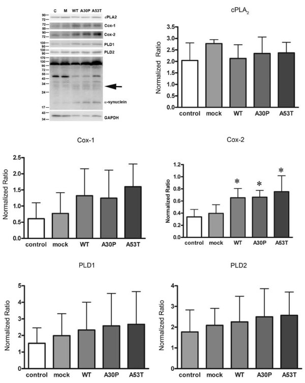
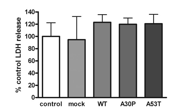
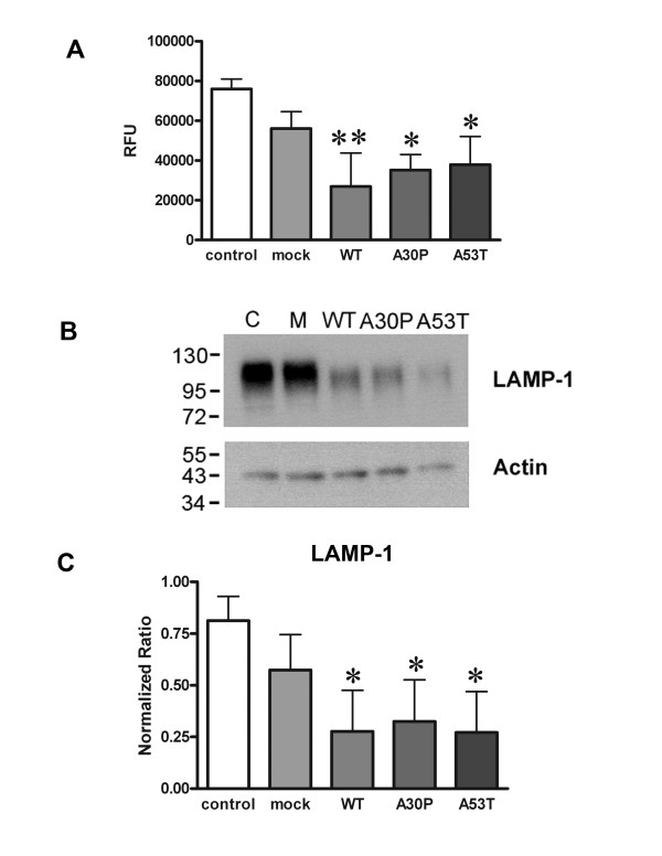
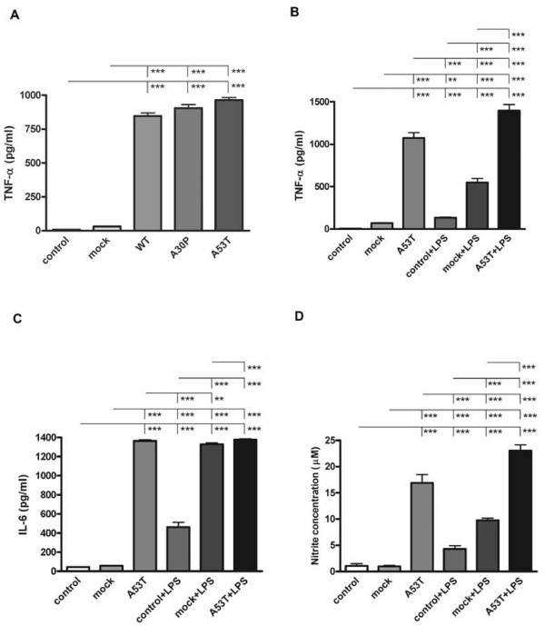
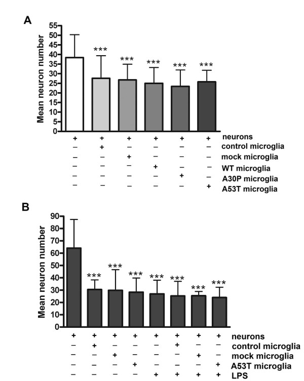
Results: As expected, over-expression of α-synuclein induced a reactive phenotype in the transfected cells. Expression of α-synuclein increased protein levels of cycloxygenase-2 (Cox-2). Transfected cells demonstrated increased secretion of the proinflammatory cytokines, tumor necrosis factor-α (TNF-α) and interleukin-6 (IL-6), as well as increased nitric oxide production. Transfected cells also had impaired phagocytic ability correlating with decreased protein levels of lysosomal-associated membrane protein 1 (LAMP-1). In spite of the increased cytokine secretion profile, the transfected cells did not exhibit increased neurotoxic ability above control non-transfected BV2 cells in neuron-microglia co-cultures.

Conclusions: These data demonstrated that over-expression of α-synuclein drives microglial cells into a form of reactive phenotype characterized by elevated levels of arachidonic acid metabolizing enzymes, cytokine secretion, and reactive nitrogen species secretion all superimposed upon impaired phagocytic potential.


Figures

Figure 1
Figure 1
α-synuclein transfected BV2 cells increased Cox-2 protein expression. BV2 cells were transiently transfected to express WT, A30P, or A53T α-synuclein for 48 hours. A) Cells were lysed and Western blotted using anti-cPLA2, anti-Cox-1, anti-Cox-2, anti-PLD1, anti-PLD2, anti- α-synuclein, or anti-GAPDH (loading control) antibodies. Protein levels of B) cPLA2, C) Cox-1, D) Cox-2, E) PLD1, and F) PLD2 were quantified and normalized to GAPDH. Graphs are the average (± SD) of five independent experiments. * p < 0.05 compared to mock transfected cells.
Figure 2
Figure 2
α-synuclein over-expression was not toxic to BV2 cells. BV2 cells were transiently transfected to express WT, A30P, or A53T α-synuclein for 48 hours. An LDH release assay was performed to determine cell viability by quantifying LDH release into the medium using commercial LDH assay reagents. Graphs are the average (± SD) of three independent experiments. Each experiment was performed with 8 replicates per condition.
Figure 3
Figure 3
Over-expression of α-synuclein attenuated the phagocytic ability of BV2 cells and lysosomal protein expression. BV2 cells were transiently transfected to express WT, A30P, or A53T α-synuclein for 48 hours. A) Transfected cells were incubated with FITC-labeled E. coli bioparticles (0.25 mg/mL) for 3 hours. After incubation, the media was removed and the signal from any unphagocytosed or membrane associated particles was quenched by incubating cells with a (0.25 mg/mL) trypan blue solution for 3 minutes. The fluorescence intensity of phagocytosed particles was measured via fluorescent plate reader (RFU). Each condition was performed with 8 replicates. B) Transfected cells were also lysed and Western blotted with anti-LAMP-1 and actin (loading control) antibodies. C) Optical density of LAMP-1 immunoreactive bands were normalized against their respective actin bands and averaged (± SD) from three independent experiments. * p < 0.05 compared to mock transfected cells, **p < 0.01 compared to mock transfected cells.
Figure 4
Figure 4
Over-expression of α-synuclein increased TNF-α, IL-6, and nitrite levels in medium from BV2 cells. BV2 cells were transiently transfected to express WT, A30P, or A53T alpha-synuclein for 48 hours with or without 25 ng/ml LPS stimulation. Media were collected and used for quantifying concentrations of secreted A) and B) TNF-α, C) IL-6 using a commercial mouse TNF-α and IL-6 ELISA. D) Media was also used to perform Griess assay to detect the levels of nitrite secreted from BV2 cells. Each condition was performed with 8 replicates. Graphs are the average (± SD) of three independent experiments. ** p < 0.01, *** p < 0.001.
Figure 5
Figure 5
Over-expression of α-synuclein did not increase neurotoxic secretion from BV2 cells. BV2 cells were transiently transfected to express WT, A30P, or A53T α-synuclein then co-cultured onto a membrane insert with 7 days in vitro mouse cortical neurons for 72 hours in the A) absence or B) presence of 25 ng/ml LPS stimulation. After 72 hours, the inserts were removed and the neurons were fixed in 4% paraformaldehyde and immunostained with an anti-MAP2 antibody. MAP2 positive cells were counted to assess viability. Experiments were performed with 8 replicates per condition. Graphs are the average (± SD) of three independent experiments. *** p < 0.001 compared to neurons only.

Similar articles

Cited by

References

    1. Kreutzberg GW. Microglia: a sensor for pathological events in the CNS. Trends Neurosci. 1996;19:312–8. doi: 10.1016/0166-2236(96)10049-7. - DOI - PubMed
    1. Kim SU, de Vellis J. Microglia in health and disease. J Neurosci Res. 2005;81:302–13. doi: 10.1002/jnr.20562. - DOI - PubMed
    1. Shaikh SB, Nicholson LF. Effects of chronic low dose rotenone treatment on human microglial cells. Mol Neurodegener. 2009;4:55. doi: 10.1186/1750-1326-4-55. - DOI - PMC - PubMed
    1. Fischer HG, Reichmann G. Brain dendritic cells and macrophages/microglia in central nervous system inflammation. J Immunol. 2001;166:2717–26. - PubMed
    1. Park JY, Paik SR, Jou I, Park SM. Microglial phagocytosis is enhanced by monomeric alpha-synuclein, not aggregated alpha-synuclein: implications for Parkinson's disease. Glia. 2008;56:1215–23. doi: 10.1002/glia.20691. - DOI - PubMed

Publication types

MeSH terms